Login
Subscribe
microscopy, techniques
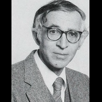
Aaron Klug, Developer of Crystallographic Electron Microscopy, Dies
Ashley P. Taylor | Nov 26, 2018 | 3 min read
The chemist and biophysicist won a Nobel prize for the development of a technique to probe the structures of nucleotide-protein complexes.

Breakthrough Prizes Recognize Aneuploidy Researcher, Biochemist
Catherine Offord | Oct 18, 2018 | 2 min read
This year’s winners also include the developers of nusinersen, an oligonucleotide therapeutic for spinal muscular atrophy.

Image of the Day: Weevil Eye
Kerry Grens | Oct 17, 2018 | 1 min read
The Nikon photomicrography competition winners of 2018 include striking close-ups of a compound eye, a fern, and an insect’s bubble house.

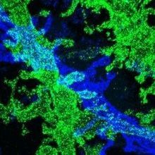
Image of the Day: Transgenic Axolotl
Kerry Grens | Oct 3, 2018 | 1 min read
Lineage tracing reveals how cells help the salamanders regrow chopped-off limbs.

Image of the Day: The Birth of a Nervous System
Jef Akst | Oct 2, 2018 | 1 min read
The winner of the 2018 Nikon Small World in Motion video competition shows the development of sensory neurons in a zebrafish embryo.

Image of the Day: Lit Up
Sukanya Charuchandra | Jul 24, 2018 | 1 min read
Researchers have updated a method to look at biochemical activities inside living cells.

Image of the Day: Eye-Opener
Sukanya Charuchandra | Jul 17, 2018 | 1 min read
An open-source microscopy method can super-resolve live cells even under low lighting.
Image of the Day: Lego Microscopy
The Scientist Staff | May 16, 2018 | 1 min read
With open-source software and Lego hardware, researchers have created a low-cost, automated method for cellular fluorescence microscopy.
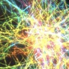
Image of the Day: Rainbotubules
The Scientist and The Scientist Staff | Apr 26, 2018 | 1 min read
By combining two microscopy techniques, researchers produce images of structures less than 10 nanometers wide.

Scientists Who Developed Cryo-Electron Microscopy Win Nobel Prize
Diana Kwon | Oct 4, 2017 | 3 min read
Chemistry Nobel goes to Jacques Dubochet, Joachim Frank, and Richard Henderson.

Image of the Day: A Swell Idea
The Scientist | Jul 19, 2017 | 1 min read
To improve the resolution of biological samples at the cellular level, researchers inflate tissues with “swellable polymers” so that they’re easier to see under the microscope.
Scientists Stretch Neurons to Image Fine Structures
Kerry Grens | Apr 18, 2017 | 1 min read
A double-expansion technique embeds brain tissue in the absorbent material of diapers to stretch out cells for easier visualization.

Live Imaging Using Light-Sheet Microscopy
Kelly Rae Chi | Nov 1, 2016 | 8 min read
How to make the most of this rapidly developing technique and a look at what's on the horizon

An Evolutionary History
Mary Beth Aberlin | Oct 1, 2016 | 3 min read
Celebrating 30 years and a resurrection

Microscopy’s Growth Through the Years
Jenny Rood | Oct 1, 2016 | 5 min read
From confocal fluorescence microscopy to super-resolution and live 3-D imaging, microscopes have changed rapidly since 1986.

Thirty Years of Progress
The Scientist | Oct 1, 2016 | 1 min read
Since The Scientist published its first issue in October 1986, life-science research has transformed from a manual and often tedious task to a high-tech, largely automated process of unprecedented efficiency.
Updated Tissue-Clearing Protocol Extends Time Frame for Imaging
Kerry Grens | Aug 23, 2016 | 1 min read
“Ultimate DISCO” uses a solvent that shrinks whole animals and preserves fluorescence for months.

Top Technical Advances 2015
Kerry Grens | Dec 23, 2015 | 5 min read
The Scientist’s choice of major improvements in imaging, optogenetics, single-cell analyses, and CRISPR

Imaging Live Tissue Without Fluorescence
Anna Azvolinsky | Oct 30, 2015 | 3 min read
Modifying a vibration-based optical technique can capture images of living tissues, researchers show.