The Scientist - Home

| 3 min read
By stamping barcodes into the genome, researchers traced cell lineages from a single starting cell to more than 1.2 million cells in a developing mouse embryo.
Miles away from the closest ocean, marine microbes thrive in the red ice and mud around Blood Falls, offering clues to microbial adaptation and past climate events.
View this Issue
Explore how blood-based biomarkers perform across Alzheimer’s disease stages, including AIBL Study findings comparing p-Tau217 and other markers against amyloid-beta and tau PET reference standards.

Learn how microvolume UV/Vis and DLS can accelerate, automate, and streamline protein developability screening while reducing sample requirements.

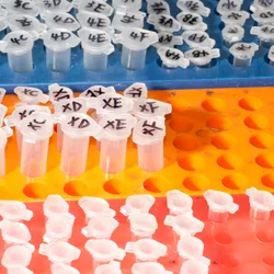
Assess protein sample purity and homogeneity in minutes with mass photometry using only nanograms of sample.


Explore how super-resolution microscopy enables particle-level characterization of lipid nanoparticle composition, cargo loading, heterogeneity, and stability, providing complementary insights for formulation development and optimization.


The placements follow the commercial launch of Pyxa at the Advances in Genome Biology and Technology (AGBT) meeting in February 2026 and mark the company's transition from Early Access into global commercial availability.
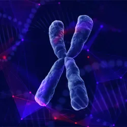
Generate robust, reproducible ATAC-seq data from as few as 1,000 nuclei. The CUTANA™ ATAC-seq Kit uses validated reagents, pre-charged Tn5 and a streamlined workflow, yielding high-quality data with less troubleshooting at a low cost per reaction.

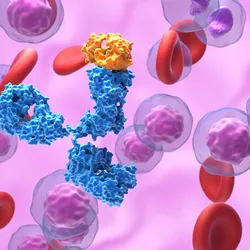
The CellRaft AIR streamlines antibody discovery with imaging, functional screening, and gentle isolation of viable single B cells and hybridomas, helping verify monoclonality and identify high producing clones faster.

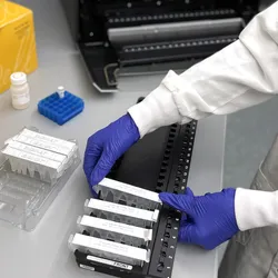
Load plasma directly into prefilled cartridges, no pretreatment, no ethanol. Under 30 minutes of instrument run time, up to 48 samples in parallel, with a high-throughput format also available.