An Inside Look at the First Optogenetics Experiment

Ed Boyden reveals how a late night in the lab and stroke of luck launched a technology that took neuroscience by storm two decades ago.

Written byDanielle Gerhard, PhD
| 2 min read
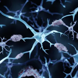
A neuron is lit up in blue and yellow against a black background.
Register for free to listen to this article
Listen with Speechify
0:00
2:00

In the early hours of August 4, 2004, Ed Boyden, now a neuroscientist at the Massachusetts Institute of Technology, sat down at the patch clamp electrophysiology rig in his graduate research advisor Richard Tsien’s lab at Stanford University. He was excited to finally test an idea that was more than four years in the making: controlling brain cells with light.

In preparation for his optogenetics experiment, he cultured rat hippocampal neurons expressing the protein channelrhodopsin-2 (ChR2), a light-gated cation channel.1 At the time, researchers weren’t sure whether they could activate ChR2 in the nonretinal cells without supplying the chemical all-trans-retinal, but Boyden intended to try it. In the presence of blue light, he hoped to see spikes emerge in the voltage recordings as an indicator of channel-driven increased neuronal activity.

Boyden secured the neurons in the microscope, placed an electrode into a cell to measure electrical currents, and flipped on the blue light. “It was kind of unknown what would happen next,” recalled Boyden. As the blue light pulsed, Boyden’s heart raced. In the very first neuron he recorded, similar to the image depicted here, he watched as spikes in neuronal activity followed the blue light with temporal fidelity. “The biggest serendipity, beyond the safety, the power, and the speed, was the fact that this chemical, for whatever weird, unknown reason, is present in mammalian neurons,” said Boyden.

Boyden ran a number of experiments that night that formed the foundation of a 2005 Nature Neuroscience paper, the result of a long-running collaboration with Karl Deisseroth, a neuroscientist at Stanford University.2 In the preceding years, Boyden and Deisseroth dreamed of developing innovative neurotechnologies for controlling brain activity. “Seeing the data that night started to make it real,” said Boyden.

  1. Nagel G, et al. Channelrhodopsin-2, a directly light-gated cation-selective membrane channel. Proc Natl Acad Sci U S A. 2003;100(24):13940-13945.
  2. Boyden ES, et al. Millisecond-timescale, genetically targeted optical control of neural activity. Nat Neurosci. 2005;8:1263-1268.
Add The Scientist as a preferred source on Google

Add The Scientist as a preferred Google source to see more of our trusted coverage.

Meet the Author

  • Black and white photo of Danielle Gerhard

    Danielle is a Senior Editor at The Scientist. She earned her PhD in psychology and behavioral neuroscience from Yale University and completed a postdoctoral fellowship in neuroscience Weill Cornell Medicine. Her research explored the cellular and molecular mechanisms of stress and depression across development. She has served as editor-in-chief of the Yale Journal of Biology and Medicine, contributed stories to BioTechniques News and Drug Discovery News, and engaged in various science outreach initiatives across Connecticut and NYC.

    View Full Profile

Related Topics

You might also be interested in...
Loading Next Article...
You might also be interested in...
Loading Next Article...
November digest cover
November 2026

Chemistry Meets Arcade Combat in “Reaction Rumble” Game

Choose your fighter! Enter the arena as chemical elements and dole out moves inspired by real-world reactions in this educational browser-based game.

View this Issue
Thumbnail

Make It Local, Make It Last: The Evolution of mRNA Protein Replacement Therapy

Engineering Bispecific Antibodies Against Emerging Viruses

Engineering Bispecific Antibodies Against Emerging Viruses

innovative-research-horizontal-lock-inverted-rgb-900px-w-72ppi
Advancing Research With Humanized Mouse Models

Advancing Research with Next-Generation Humanized Mouse Models

bioxcell
3d illustration showing amyloid plaques in Alzheimer's disease

Best Practices for Blood-Based Amyloid Pathology Testing

fujirebio logo

Products

Abstract image of circles and concentric lines on a blue and pink background.

MX Analytical and Precision Balances: Effortless Weighing and Secure Data Handling

Mettler
3D multicolored conceptual image of antibody molecules

Premium BsAb: LC-MS-Assisted Bispecific Antibody Production with >98% Heterodimer Purity

WuXi Biologics
Abstract conceptual image of AI technology for medical science research.

Exploring a New Era of Pharmaceutical Innovation

A photograph of plastic fragments floating in water, symbolizing contamination from sample storage tubes.

LVL SAFE® 2D-Coded Tubes: The Perfect Tube for Your Sample

AutoGen