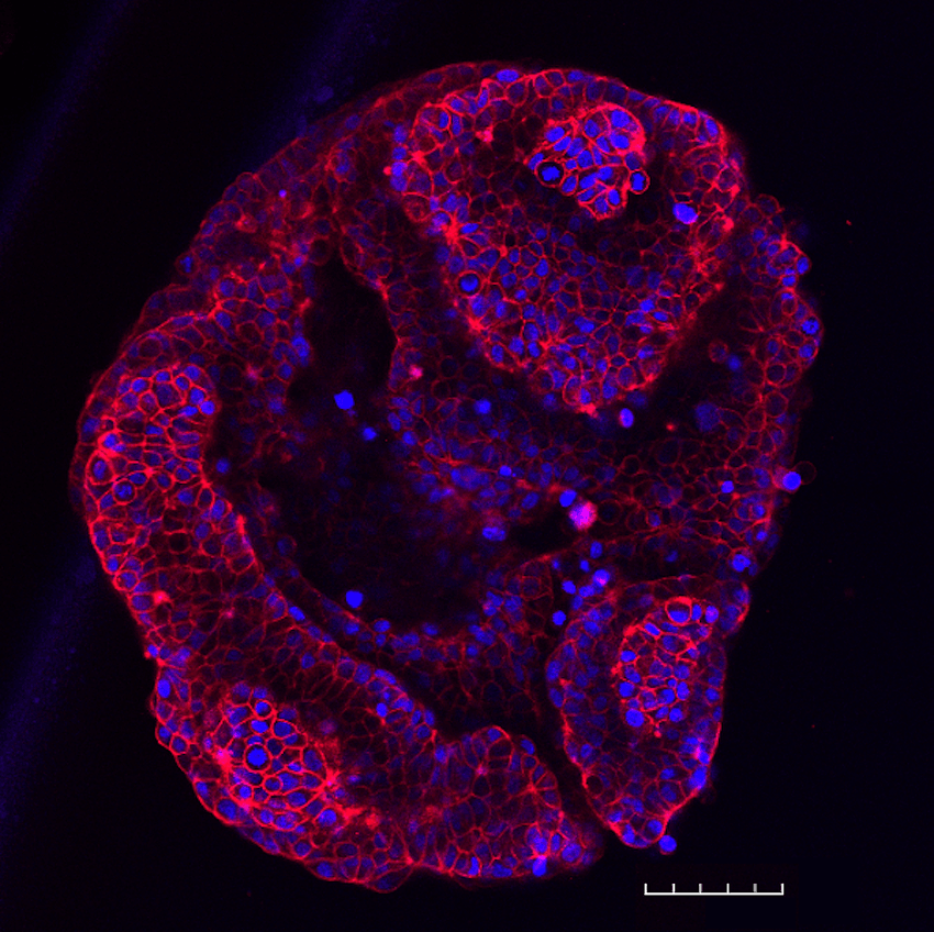
T47-D spheroid stained with NucSpot® Live 650 (nuclei, blue) and CellBrite® Steady 594 (membranes, red). T47-D cells were plated at 5,000 cells per well in a LICORbio 3D Essentials Kit ULA 96-well plate and cultured for 48 hours. The spheroid was stained with dyes in complete medium for 30 minutes without a wash step and imaged on an Evident FV4000 confocal microscope with 20X objective to create a tile-stitched 3D projection image; scale bar: 100 um.
Unlike conventional membrane dyes, such as lipophilic carbocyanine dyes (i.e. DiI or DiO) or PKH Dyes, that are rapidly internalized by endocytosis, CellBrite® Steady Membrane Staining Kits deliver stable, long-term cell surface staining for live cell imaging. Signal is retained at the plasma membrane for 24 hours or longer, with validated performance for up to several days in culture. This durability comes from the unique chemistry of CellBrite® Steady Dyes, fluorescent membrane probes that distribute between the cell surface and intracellular compartments so that cell boundaries remain clearly defined even during extended time-lapse and longitudinal imaging experiments.
Each kit includes CellBrite® Steady Enhancer, an optional reagent that masks intracellular fluorescence for more selective visualization of cell surface membranes and sharper boundary contrast. Researchers can choose from nine dye colors spanning blue to near-IR, including STORM-compatible options, making CellBrite® Steady Dyes flexible for multiplexed fluorescence imaging, confocal microscopy, and super-resolution applications.
Features:
- Retained on cell surface, unlike other membrane dyes that are rapidly internalized
- Image live cell surface membranes for 24 hours or longer
- Image in your preferred channel with 9 color options from blue to near-IR
- Optional Enhancer masks intracellular signal for selective imaging of cell surface
- Rapid, even staining in complete cell culture medium
- Dye colors from blue to near-IR, with STORM-compatible options
CellBrite® Steady Dyes stain rapidly and evenly in complete cell culture medium, with no wash step required for confocal imaging. The dyes exhibit low toxicity and are compatible with continuous incubation in culture medium throughout the imaging timeline. The staining is ideal for live-cell applications where membrane integrity and cell viability over time are essential. Enhancer may be added after staining or co-incubated with the membrane dye, offering flexibility to minimize any potential cytotoxic effects in sensitive cell types.
Product Specifications:
| Specification | Value |
| Probe cellular localization | Cytoplasmic membrane/cell surface |
| For Live or Fixed Cells | For live/intact cells |
| Assay type/options | long term staining |
| Detection method/readout | microscopy |
| Fixation options | Fixation not recommended |
| Colors | Blue, Green, Red, Far-red, Near-infrared |