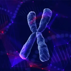
Telomeres are structures at the ends of chromosomes that protect them from degrading or interacting with other pieces of the genome, which can lead to accelerated aging and cancer. While 80 to 95 percent of cancers maintain their “immortality” by using the enzyme telomerase to maintain the length of their telomeres, a smaller subset—predominantly soft tissue and bone cancers—rely on the alternative lengthening of telomeres (ALT) pathway.
The ALT pathway is thought to occur as a secondary, last-ditch survival mechanism for cancer cells. Instead of using telomerase to maintain telomeres, this process relies on DNA repair pathways to elongate the telomeres and makes them more resistant to standard therapies.
More than a decade ago, Roderick O’Sullivan, a geneticist at the University of Pittsburgh Medical Center, investigated how telomeres in ALT-positive cells differed from those expressing telomerase using a proteomic screen. What he found stumped him—he saw centromeric proteins in the ALT telomeres. An observation which struck him as odd because centromeres and telomeres are considered distinct both in location and function; consequently, he discarded the finding.

O’Sullivan and Bhargava are excited to further investigate the extent of their ALT findings.
University of Pittsburgh Medical Center
However, Ragini Bhargava, a postdoctoral researcher in O’Sullivan’s group, saw the same strange sight several years later. This motivated them to revisit the observation and dive deeper. In a new study, published in Nature, O’Sullivan and his colleagues found that centromeres left footprints on telomeres in ALT tumors.1 These interactions left discrete marks that preserved the cancer cells’ telomere integrity. Currently, there is not a good diagnostic for detecting ALT tumors, which are highly aggressive; thus, this genomic signature is an attractive target that researchers could potentially use as a biomarker.
When O’Sullivan initially disregarded the centromere proteins in telomeres as an artifact, Bhargava pressed on. Using immunostaining in a standard ALT cell line, she found overlapping telomere and centromere-associated motifs, called alpha satellite repeats, which typically contain centromere proteins A and B (CENP-A, CENP-B). She confirmed this finding with further DNA sequencing. O’Sullivan added, “There was a clear association and presence of that motif in telomeres from every ALT cancer line selected.” They observed the same motifs in ALT neuroblastoma samples, demonstrating that this phenomenon also occurred in human cancers in vivo.
To further characterize this molecular signature, they then used directed methylation with long-read sequencing. Here, the team identified discrete footprints of CENP-A chromatin assembled at telomeric locations on subsets of chromosomes. The researchers saw CENP-A footprints on some chromosomes, positioned precisely where acute loss of DNA methylation occurred. This method aided the researchers in mapping the distinct epigenetic changes associated with ALT tumor cells.

The researchers observed a centromere protein “lighting up” at telomeres, which led them to dig deeper.
University of Pittsburgh Medical Center
To better understand the process that contributes to these centromeric signatures, the researchers modeled ALT activation by disrupting alpha-thalassemia intellectual disability X-linked (ATRX) protein expression. ATRX is the most frequently altered gene in ALT cells. When the team compared ATRX knockout, ALT-positive cells with control non-cancer cells, they saw that ATRX loss and DNA hypomethylation facilitated the acquisition of these centromeric chromatin signatures. Meanwhile, disruption of CENP-A accumulation affected telomere integrity, underscoring the centromeric protein’s role in maintaining productive ALT cancer. Together, these findings provide evidence for telomere-centromere interactions and that these footprints play a role in ALT tumors.
“[This work] really does feel like a right place at the right time kind of project,” Bhargava said. “I feel like if we did the same study five years ago, it would have looked different.” She added that advances in sequencing technologies and telomere-to-telomere assembly played immense roles in making the analysis for the project possible.
“[This paper] is really lovely. The strengths of it are the wide range of cutting-edge genomic techniques that they've used and the number of cellular systems that they've used,” said Hilda Pickett, a geneticist at the University of Sydney and Children’s Medical Research Institute, who was not involved in the study. Pickett studies telomere maintenance mechanisms and has had a long-standing interest in ALT. “I'm fascinated that we've not really acknowledged or recognized it before. So, I think the work done has been so comprehensive,” she said.
For what this means for the field, Pickett is interested in the extent of these molecular footprints, such as why they localize to those areas at the telomeres and whether this phenomenon is conserved in other cancers, and how insights from this work may eventually lead to developing therapeutics. “[It’s] a very powerful system going forward,” she added.
O’Sullivan and Bhargava echoed these sentiments. While cancer cells are devious and will find ways to adapt, the team hopes that further studies will reveal a targetable vulnerability in these cells.
- Bhargava R, et al. Genomic and epigenomic centromeric footprints preserve telomere integrity in ALT cancers. Nature. 2026.