Tattoos are a longstanding form of self-expression through body art but given their nature of breaking the skin to deposit ink, the safety of the products that parlors use is imperative. While temporary inflammation is normal, in 2012, the Food and Drug Administration (FDA) helped investigate a series of reported severe rashes in people after a tattoo application. The cause turned out to be an ink contaminated with a species of non-tuberculosis mycobacteria (NTM).1 These and other incidences led to product recalls predominantly due to microbial contamination.2
“As a result of that, we started to look into what did we need to do from a regulatory perspective to make sure that these products were safe and being safely used,” said Linda Katz, the director for the Office of Cosmetics and Colors at the FDA, which regulates tattoo and permanent makeup inks.
In their follow up studies, Katz and others from the agency identified contamination with aerobic bacteria in one-third of all the inks assessed between 2015 and 2019.3-5 In a recent study, Katz and her colleagues expanded their investigation to assess contamination with anaerobic microbes. The team identified anaerobic bacteria in addition to aerobic microbes in several tattoo and permanent makeup inks.6 The findings, published in Applied Environmental Microbiology, highlight the need for continued monitoring to ensure the products’ safety.
“When you think about tattoo pigment, which is meant to be [in] a place where it probably doesn't belong forever, if there is contamination, especially with an anaerobe versus an aerobe…anaerobes are much harder for our immune system to combat,” said Adam Friedman, a dermatologist at George Washington University who was not involved with the study. “What's unique about it is that they don't just focus on [NTM] which is usually what the spotlight’s on. They're actually looking at a range of bacteria, both aerobic and anaerobic,” said Friedman.
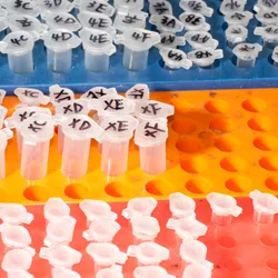
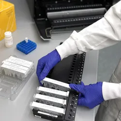
For their experiments, the FDA scientists sampled 75 different inks from multiple manufacturers that sold products in the United States by swabbing culture plates for growth in aerobic, anaerobic, and low oxygen conditions. They recovered bacteria from 26 inks. Most products only contained one isolate, although some contained two.
The researchers performed 16S ribosomal RNA to identify the species responsible for the ink contamination. They identified 22 distinct species across all the contaminated products, including two anaerobic species. Eight species are potential pathogens. The most common taxa overall were Staphylococcus species, and all were considered potential pathogens.
“Because of the continued presence that we've been finding of microbial contamination, we're continuing to inspect and to look at these products further,” Katz said. According to the researchers, the recovery of microbes from tattoo and permanent makeup inks, some of which were labeled sterile, prompts further study into the manufacturers’ sterilization methods.
“We need to ensure that those receiving [tattoos] are safe and those who are doing [tattoos] are also safe in the sense of they are not put in potentially liability harm’s way in terms of using contaminated ink,” Friedman said. “This is a very long-standing industry that really deserves the appropriate quality assurance.”
- Kennedy BS, et al. Outbreak of Mycobacterium chelonae infection associated with tattoo ink. New Engl J Med. 2012;367(11):1020-1024.
- Yoon S, et al. Recalls of tattoo and permanent makeup inks in the United States and a follow-up microbiological survey of inks with a previous recall history. Front Public Health. 2023;11:1279884.
- Nho SW, et al. Microbial contamination of tattoo and permanent makeup inks marketed in the US: A follow-up study. Lett Appl Microbiol. 2020;71(4):351-358.
- Nho SW, et al. Microbiological survey of commercial tattoo and permanent makeup inks available in the United States. J Appl Microbiol. 2018;124(5):1294-1302.
- Yoon S, et al. Microbiological survey of 47 permanent makeup inks available in the United States. Microorganisms. 2022;10(4):820.
- Yoon S, et al. Detection of anaerobic and aerobic bacteria from commercial tattoo and permanent makeup inks. Appl Environ Microbiol. 2024;90(7):00276-24.