When a newborn trades the warmth of the womb for a breath of air and lets out a cry, everyone in the delivery room heaves a sigh of relief. But if a baby struggles to breathe, those moments can become a race against time.
Oxygen deprivation during birth can cause neurological complications or be fatal, but the currently-used fetal electronic monitor—which tracks the baby’s heartbeat—does not directly indicate fetal hypoxic distress.
“What's really striking is that, despite all of the advances that we've made in medicine, we're still relying on electronic fetal heart rate monitoring, which has been around for over 50 years,” said Sandy Elmehrath, a women’s health researcher at the Wyss Institute at Harvard University (the Wyss Institute).
An unreliable tool during the critical minutes at the time of delivery means doctors have to play a high-stakes guessing game about whether a newborn is suffocating. “This uncertainty can go both ways,” explained Elmehrath, wherein clinicians end up performing more than 80,000 unnecessary cesarean sections resulting in one billion dollars in avoidable healthcare costs. On the other hand, more than 130,000 babies are born each year with severe hypoxia.

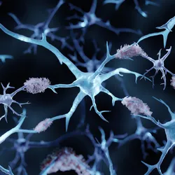
During pregnancy, the placenta releases extracellular vesicles into the maternal bloodstream. These vesicles carry molecular cargo, including proteins and microRNAs, that can provide information about what’s happening at the maternal-fetal interface.
Silvi Lybbert, Wyss Institute
To solve this problem, Elmehrath joined forces with Peter Nguyen, a synthetic biologist and protein engineer at the Wyss Institute. Together, they turned toward extracellular vesicles (EVs)—nanosized, membranous structures secreted into the extracellular space—and conceptualized a project called Fetal Extracellular Vesicle Monitoring of Oxygenation (FEMOx) to quantify hypoxia-associated biomarkers in maternal blood samples. This molecular diagnostic technology will offer a strategy to objectively and non-invasively track fetal oxygen levels, thus “taking the guesswork out of fetal hypoxia,” said Nguyen.
Decoding Molecular Messages from the Placenta
Elmehrath and Nguyen focused on EVs because scientists have increasingly found that these structures are a promising source of circulating biomarkers for diagnosing a number of conditions.1
“You could think of [EVs] as little messages in a bottle that cells send to one another, [like] little packets of information,” explained Nguyen. “It is this communication network that we can actually eavesdrop in on and figure out what's going on with the cells.”
Fetal cells facing oxygen deprivation send out EVs to maternal blood which contain cargo such as RNA or proteins representing hypoxic stress. The researchers aim to quantify hypoxia-associated microRNAs in placental EVs circulating in the mother’s body.
But to do this, they have to ensure that they measure placenta-specific EVs in maternal blood. “[EVs] actually have zip codes to them,” said Nguyen, so researchers can tell which tissue the EVs have originated from. “If you know the marker—which we do know the marker for placental EVs—you can actually pluck them out of the blood and extract those specific EVs so that you can read those messages.”
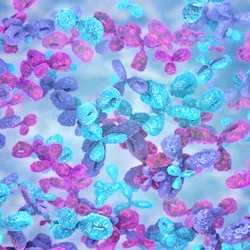
At the moment, Elmehrath, Nguyen, and their team are working on optimizing a way to obtain placental EVs from maternal blood samples. They will rely on a multilayer microfluidic chip developed at the Wyss Institute, which uses antibody arrays to isolate EVs from the blood. Then, they plan to carry out RNA sequencing to identify microRNAs differentially expressed in samples from hypoxic and healthy conditions. This will help them build a microRNA biomarker signature panel for fetal oxygen distress.
Translating Molecular Insights into Safer Childbirth

FEMOx can help in bedside assessment of fetal hypoxia by EV analysis of maternal blood.
Partially generated through AI, Wyss Institute
However, both Elmehrath and Nguyen admitted that this path will not be straightforward or easy. One of the biggest challenges they anticipate along the way is identifying biomarkers that can sensitively indicate fetal hypoxia, said Elmehrath. “We're trying to detect a very small amount of placental EVs and biomarkers from maternal blood,” she said.
Nguyen added that a majority of EVs do not carry any signals, which makes it difficult to identify hypoxia-associated biomarkers in EVs with certainty. But he noted that the EV fraction in maternal blood spikes significantly during the third trimester, which could provide a window for detecting the biomarkers.
Recognizing the promise of this work to address a critical unmet need in maternal-fetal care, the researchers applied for and recently bagged a contract from the US federal funding agency Advanced Research Projects Agency for Health. “This isn't just a science project,” said Nguyen, noting that the team is already considering regulatory and commercialization strategies.
“The goal is not to just develop this exciting technology, there's really a strong emphasis on translating it into something that could really reach patients and be used in real clinical workflows,” said Elmehrath.
- Di Bella MA, Taverna S. Extracellular vesicles: Diagnostic and therapeutic applications in cancer. Biology (Basel). 2024;13(9):716.