Shingles can cause pain that is notoriously difficult to manage and can last for months to years for some people.1 The pain can be so severe that it causes a significant decline in quality of life and can be accompanied by suicidal thoughts and emotional distress.2
If the pain lasts longer than three months, doctors call the condition post-herpetic neuralgia.3 Because the underlying mechanisms driving this persistent pain are unknown, treatment options are centered on relieving symptoms but largely fall short. Less than 50 percent of patients achieve meaningful pain relief.4
However, new research from my team published in the journal Annals of Neurology uncovered that microparticles circulating in the blood called exosomes may provide an explanation for some of the most puzzling features of this neuropathic pain condition as well as new targets that could lead to new treatments.5,6
Chickenpox Reawakens As Shingles
The varicella zoster virus that causes chickenpox has infected over 90 percent of the world’s population.3 Most people contract the virus during early childhood in temperate regions, but in tropical regions, initial infection typically occurs later in adolescence and early adulthood.
After this first infection, the virus enters a state of dormancy within pain-sensing neurons. Even children vaccinated for varicella, such as those in the United States since 1995, still have dormant virus in their neurons, since the vaccine uses a live but weakened strain.
In approximately a third of the world’s population, the virus will reactivate decades later to cause the infamously painful shingles rash; for reasons yet unknown, rates of shingles are increasing worldwide.3 For most people, the pain will resolve in a couple of weeks. However, about 10 percent to 18 percent of patients will progress to post-herpetic neuralgia months after the rash has cleared. In some cases, the pain can persist for years or the rest of their lives.
While anyone who has shingles is at risk of developing post-herpetic neuralgia, the risk rises significantly with age.4
Exosomes And Irritated Neurons
Researchers and clinicians do not fully understand what causes post-herpetic neuralgia. In patients with post-herpetic neuralgia, while the shingles rash has cleared with seemingly normal-looking skin, skin biopsies show a puzzling reduction in sensory nerve fibers in the painful areas.7 Counterintuitively, this decrease in nerve fibers in the skin can lead to heightened pain in many patients. What prevents these nerve fibers from repairing after the infection has cleared is unknown.
Researchers have proposed that ongoing or intermittent viral replication within sensory neurons is likely continuing to damage or kill these cells months after the virus reactivates. However, antiviral treatment that shuts down viral replication does not reliably prevent or reduce post-herpetic neuralgia.8 Thus, neurovirologists like me have been looking for noninfectious contributors to this condition.

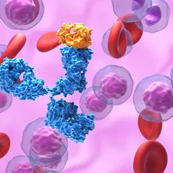
In our newly published research, my team and I investigated the role that noninfectious microscopic particles circulating in the blood called exosomes may play in the development of post-herpetic neuralgia.5,6 Exosomes are released from every type of cell in the body. They carry bioactive cargo – material such as proteins and nucleic acids that can change a recipient cell’s behavior or specific cellular process – and shuttle them from one cell to another. This process helps cells communicate with each other and is essential for normal bodily functions.
My laboratory previously discovered that exosomes in the blood of patients with an active shingles rash can increase stroke risk and inflammation. Thus, we hypothesized that circulating exosomes in the blood of post-herpetic neuralgia patients may also be contributing to their severe pain.
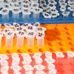
My team and I isolated exosomes from the blood of seven patients with post-herpetic neuralgia and compared them to those in the blood of seven patients without the condition.5 The contents of the exosomes of those with post-herpetic neuralgia had significantly higher concentrations of proteins known to suppress the growth of neurons. Did this finding mean that patients with post-herpetic neuralgia have particles in their blood that are preventing their pain sensory neurons from fully regenerating?

To test this assumption, we exposed human sensory neurons in a petri dish to exosomes isolated from the blood of people with or without post-herpetic neuralgia. Using live-cell imaging, we tracked and measured the ability of these neurons to extend and interact with each other in the petri dish. As suspected, neurons exposed to post-herpetic neuralgia exosomes were significantly stunted and unable to form a strong network with other neurons.5 In comparison, neurons exposed to the exosomes of people without the condition saw no measurable disruptions to their function.
When we measured the genetic activity of pain sensory neurons following exposure to post-herpetic neuralgia exosomes, we found that these neurons underwent changes that actively prevent the formation of growth cones – structures neurons need to grow and repair themselves.9 Furthermore, we found evidence that post-herpetic neuralgia exosomes keep pain-sensing neurons in an overactive state, rendering them hypersensitive to pain signaling.

Future of Neuropathy Treatment
Our findings suggest that targeting the underlying cause behind nerve irritation and failed regeneration could help lead to more effective treatments for post-herpetic neuralgia by targeting the source of the pain.
Furthermore, my team and I believe this phenomenon is not unique to post-herpetic neuralgia. It could likely extend to other painful neuropathies, such as diabetic neuropathy, which is also associated with failed nerve regeneration.10
Currently, my team is collecting sequential blood samples for up to a year from patients with shingles who later develop post-herpetic neuralgia, as well as from patients with shingles that resolves with no lingering pain. This comparison will be crucial to determine specifically what cargo exosomes are carrying that contribute to this debilitating chronic pain condition.
Andrew Bubak, Associate Professor of Neurology, University of Colorado Anschutz
This article is republished from The Conversation under a Creative Commons license. Read the original article.
- Adriaansen EJM, et al. 8. Herpes zoster and post herpetic neuralgia. Pain Pract. 2025;25:e13423.
- van Wijck AJM, Aerssens YR. Pain, itch, quality of life, and costs after herpes zoster. Pain Pract. 2017;17:738-746.
- Bubak AN, et al. Varicella zoster virus infection. Nat Rev Dis Primers. 2026;12:58.
- Johnson RW, Rice ASC. Postherpetic Neuralgia. N Engl J Med. 2014;371(16):1526-1533.
- Niemeyer CS, et al. Circulating exosomes drive persistent neuronal dysfunction in post-herpetic neuralgia patients. Ann Neurol. 2026.
- Kalluri R, LeBleu VS. The biology, function, and biomedical applications of exosomes. Science. 2020;367:eaau6977.
- Oaklander AL. The density of remaining nerve endings in human skin with and without postherpetic neuralgia after shingles. Pain. 2001;92(2):139-145.
- Chen N, et al. Antiviral treatment for preventing postherpetic neuralgia. Cochrane Database of Systematic Reviews. 2014;2.
- Igarashi M. Molecular basis of the functions of the mammalian neuronal growth cone revealed using new methods. Proc Japan Acad Series B. 2019;95(7):358-377.
- Truini A, et al. Does the epidermal nerve fibre density measured by skin biopsy in patients with peripheral neuropathies correlate with neuropathic pain? Pain. 2014;155(4):828-832.