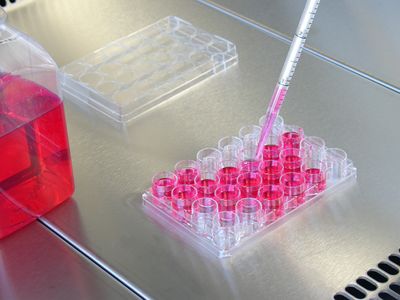
ABOVE: GlycoRNAs join glycolipids and glycoproteins as part of the cell's sugar-coating.
RYAN FLYNN
The emergence of nucleic acids and that of proteins have sometimes been called the first and second evolution revolutions, as they made life as we know it possible. Some experts argue that glycosylation—the addition of glycans to other biopolymers—should be considered the third, because it allowed cells to build countless molecular forms from the same DNA blueprints. It’s long been believed that only proteins and lipids receive these carbohydrate constructs, but a May 17 paper in Cell that builds upon a 2019 bioRxiv preprint posits that RNAs can be glycosylated, too, and these sugar-coated nucleic acids seem to localize to cell membranes.
Anna-Marie Fairhurst, who studies autoimmunity at the Agency for Science, Technology and Research in Singapore, describes the study as exciting. “Obviously, it’s the first time ever that we’ve seen this with RNA,” she says, adding that the diversity of methods used to demonstrate the presence of glycoRNAs makes the findings especially robust.
What really intrigues her are the parts present in the 2021 Cell paper that aren’t in the 2019 preprint—in particular, that glycoRNAs appear to predominantly end up on the cell’s outer membrane. There, they can attach to two kinds of sialic acid-binding immunoglobulin-type lectins (Siglecs)—a family of immune receptors implicated in several diseases, including systemic lupus erythematosus (SLE). All of this suggests glycoRNAs may play a role in immune signaling. “It’s a really exciting era of science,” Fairhurst says.
Going against glycobiology dogma
Ryan Flynn, the first author on the new paper and an RNA biologist at Harvard University and Boston Children’s Hospital, says he made the startling discovery of glycoRNAs while working in chemical biologist Carolyn Bertozzi’s lab at Stanford University. Bertozzi says she was skeptical at first but came around after thinking about how her own assumptions might be shaping her views. “We bring to every experiment all this unconscious bias,” she explains, and once she re-examined her own, she found no reason to think glycoRNAs shouldn’t exist. “These are ancient molecules,” she says. “There’s no reason to just presume that they wouldn’t have found a way to connect and to create new biology.”
These are ancient molecules . . . There’s no reason to just presume that they wouldn’t have found a way to connect and to create new biology.
—Carolyn Bertozzi, Stanford University
As it happens, Flynn did set out to overturn glycosylation dogma when he joined Bertozzi’s lab as a postdoc in 2017—although it didn’t happen the way he expected. At first, he explains, he had his eye on a quirky cytosolic protein glycosylation pathway because he’d noticed that one of its key enzymes has an RNA-binding domain. If there’s a glycosylation enzyme with the potential to bind RNA, and it’s functioning in the cytosol where RNAs tend to be, he reasoned, it could be sticking sugars to RNAs, too.
To search for the existence of these structures, “it was really important that I had access to things that were not dependent on high temperatures, and not dependent on metals that might otherwise degrade the RNA,” he says, and that’s exactly what Bertozzi’s lab had to offer. She’s a pioneer in the field of bioorthogonal chemistry, which aims to develop chemical methods for tracking biomolecules in their native environments. Her lab was brimming with reagents that label specific kinds of glycans without harming other molecules or setting off side-reactions.
See “Carolyn Bertozzi: Glycan Chemist”
Flynn set to work adding these glycan-labeling compounds to HeLa cells and then isolating RNA from them to see if any glycan signal remained after he’d removed all proteins and lipids. He says he thought he might see a signal when he labeled the kind of glycans used in that cytosolic glycosylation pathway.
However, months of experiments failed to support that hypothesis.
Instead, something strange kept happening with what was supposed to be a negative control: cells treated with ManNAz, an azide-labeled precursor for sialoglycans, a group of glycans known for their role as modifiers of secretory and cell surface proteins and lipids. Once the cells were given the chance to incorporate ManNAz, they were lysed with TRIzol, which breaks apart cellular components without damaging RNAs, and any surviving proteins were chopped up with proteases. The idea was that there’d be no azide signal at the end, as sialoglycans are attached to proteins and lipids in the endoplasmic reticulum and Golgi, where RNAs have no business being. “I was like, there’s no way that a reagent that labels sialoglycans is going to end up labeling an RNA, even a glycoRNA,” Bertozzi says, but those experiments consistently gave Flynn positive signals.
So, the team dug further. Not only did the glycoRNAs the team found contain this specific subgroup of glycans, they appeared to largely consist of YRNAs, a family of small, highly conserved noncoding RNAs whose cellular functions remain unclear, although previous studies have suggested they may play a role in oncogenesis and autoimmunity. The specificity of both the glycans and the type of RNAs involved strongly point to their being attached to one another with an enzyme, says Bertozzi.
See “Glycans May Bind to RNA, Initial Findings Suggest”
Furthermore, once the researchers started looking for them, they found these glycoRNAs in numerous established cell lines, including cancer-derived ones such as HeLa and T-ALL 4118 cells, as well as stem cell–derived CHO and H9 cells. They were even able to detect glycoRNAs in liver and spleen cells extracted from live mice that received intraperitoneal injections of ManNAz, suggesting that glycoRNAs are everywhere.
By 2019, the team members felt they had enough supportive data to submit their findings, so they put the preprint version up on bioRxiv. It made a splash in the scientific community, but without peer review, some remained skeptical. Now, after even more experiments and a rigorous review process, the team says its data have become even more compelling.
GlycoRNAs decorate cells
“They clearly have isolated a covalent RNA-glycan conjugate,” says Laura Kiessling, a chemical biology researcher who studies carbohydrates at MIT and was not involved in the study. However, big questions remain, including what these glycoRNAs do and how they form. For instance, it’s unclear exactly how the RNAs and glycans are physically connected to one another, she notes, and without that information, she’s not quite convinced that the binding happens enzymatically.
Flynn and Bertozzi suggest that the RNAs are glycosylated much in the same way proteins are, and that it even requires some of the same proteins. As noted in the original preprint, when they inhibited key enzymes involved in glycosylation, glycoRNAs disappeared in a dose-dependent manner. Similarly, cell lines engineered to have errors in protein glycosylation produced very little glycoRNA. But for RNAs to be glycosylated by the same pathway as proteins “would be weird,” Kiessling says, noting that multiple glycosylation steps only proceed after a check for proper protein folding. “It’s hard for me to imagine exactly how that would occur with RNA.”
The researchers were even able to detect glycoRNAs in liver and spleen cells extracted from live mice, suggesting that glycoRNAs are everywhere.
Fairhurst says she also wants to know more about the synthesis pathway. She has lots of other questions, too, which she says is a good sign. “A really good, exciting paper leaves a lot more questions than it does answers,” she notes.
While the 2019 preprint raised many of these questions, some are unique to the new data presented in the Cell version. Perhaps the biggest addition to the work was the discovery of where these glycoRNAs spend their time—stuck on the outsides of cells, explains Flynn. The team demonstrated this by briefly exposing some ManNAz-labeled HeLa cells to an enzyme that can cleave sialic acid glycans from the cell surface. If the glycoRNAs were on the outside, they would be cut off, and the total amount of glycoRNAs remaining would drop. And that’s exactly what they found: the glycoRNA signal started to decrease after as little as 20 minutes of incubation with the sialidase and was reduced by more than 50 percent after an hour, which the team suggests means that more than half of a cell’s glycoRNAs are stuck on its outer membrane.
The researchers further probed the hypothesis of extracellular localization by labeling living cells with an antibody that binds to double-stranded RNA. About one-fifth of a culture of HeLa cells were positive for antibody staining, and the label was sensitive to RNase treatment, further supporting the idea that glycoRNAs are indeed present on the outer cell membrane. “That opens up a lot of ideas, and a lot of possibilities, functionally and mechanistically, for what they could be doing,” says Flynn.
One of those possibilities is that glycoRNAs are involved in cell-to-cell signaling, especially in an immune context, as that’s a known function of membrane glycolipids and glycoproteins. Bertozzi had already been investigating the ligands of Siglecs, a group of sugar-binding receptors that modulate immune reactions, so the team decided to see if any of them bound to glycoRNAs. They first treated HeLa cells with different Siglecs to show that the receptors bound normally, then treated the cells with RNase. Lo and behold, the binding of Siglec-11 and Siglec-14 dropped precipitously, suggesting that their ligands were cleaved from the surface by the RNA-cutting enzyme.
Bertozzi says the experiment indicated glycoRNAs are ligands for Siglec-11 and Siglec-14, and if so, they’d be the first identified for Siglec-11.
“As a receptor family, [Siglecs have] kind of been ignored,” notes Fairhurst, so the fact that these glycoRNAs can interact with them is very exciting, she says. “My immediate desire is to see whether they are associated with diseases, particularly in SLE,” she adds.
GlycoRNAs and disease?
Lan Lin, an RNA biologist at the University of Pennsylvania and the Children’s Hospital of Philadelphia, says she found the 2019 preprint so interesting that she applied for and received a pilot grant from the Frontiers in Congenital Disorders of Glycosylation (CDG) Consortium to study the roles glycoRNAs may play in CDG, a group of rare congenital conditions arising from mutations in protein glycosylation pathways. Because RNA glycosylation may be related to protein glycosylation, she tells The Scientist, “it was only rational or reasonable for [my colleagues and I] to hypothesize that . . . some of these patients might have differences in the glycoRNA in their system,” and therefore, CDG conditions could be used to examine the potential functions of glycoRNAs.
So far, she says, her team hasn’t detected any consistent differences in glycoRNAs between the cells of healthy controls and CDG patients. She says that may be because differences are more qualitative than quantitative, such as alterations to the sugars themselves or the subset of RNAs that are glycosylated. Alternatively, she notes, the new data in the 2021 Cell paper may provide an explanation: the membrane localization of glycoRNAs wasn’t in the preprint, so “maybe we are looking in the wrong place,” she muses.
It’s also possible that new methods are needed to detect glycoRNA differences between cells. She points out that a major limitation of the study is that the ManNAz labeling method can’t readily be applied to preserved human tissue samples or blood samples.
Fairhurst says she’d like to see more work in primary cell cultures rather than immortalized ones, especially leukocyte subtypes, where one might expect pronounced differences if the RNAs have a role in immunity. For example, she says she’d like to see whether, in people with conditions like SLE, different cell types have fewer or more glycoRNAs, though “obviously, those experiments are really challenging.”
Seeing these big milestones is amazing
—Anna-Marie Fairhurst, Agency for Science, Technology and Research in Singapore
Still, she says, “seeing these big milestones is amazing.”
Kiessling says she thinks glycoRNAs could be “really important” in the field of glycobiology. Her lab focuses on how carbohydrate-binding proteins can “read” glycans on the surfaces of cells, she explains, so these glycoRNAs could be “a new kind of information to read.” Lin points out that the findings are especially impactful for RNA researchers, as they suggest a whole new kind of post-transcriptional modification in need of investigation. Because glycoRNA sits at the intersection of glycobiology, immunology, and RNA biology, says Bertozzi, “Ryan’s discovery has brought together these disparate worlds.”
Flynn and Bertozzi say they’re hoping to start answering some of the many questions that remain, including how the glycans attach to RNAs and how and where that happens. The most exciting part, they say, will be the investigations into what glycoRNAs do.
R. Flynn et al., “Small RNAs are modified with N-glycans and displayed on the surface of living cells,” Cell, doi:10.1016/j.cell.2021.04.023, 2021.


