Live-cell imaging

Red, green, and blue fluorescently labeled skin cells on a black background.

In Living Color: Exploring Advances in Quantitative Live Cell Imaging 

Fluorescence microscopy image of fibroblasts in culture.

Channeling Success: Simultaneous Multicolor Live Imaging

Microscopic image of a live amoeba.

Illuminating Specimens Through Live Cell Imaging

Conceptual 3D image of glowing neurons on a dark background depicting electrical activity and synaptic connections.

Measuring Long-Term Neuronal Activity in Cell Culture 

An image of neurons, astrocytes, and other glial cells acquired using fluorescence microscopy.

Troubleshooting Fluorescence Microscopy Experiments

An illustration of a dividing cancer cell.

Striking a Balance for Perfect Images

Visualizing apoptosis in a DU-145 cancer cell

Observing Cells in Their Natural State with Digital Holographic Cytometry

Fluorescence microscopy image of cells expressing fluorescent biosensors. Green and magenta fluorescence is observed outside of the cell nuclei.

Choosing Fluorescent Reagents for Every Live Cell Application

Whole-Well Brightfield Cell Counting with the Latest in Microplate Imaging Technology 

Whole-Well Brightfield Cell Counting with the Latest in Microplate Imaging Technology

Characterizing CAR T Cells

Phenotypic and Functional Characterization of CAR T Cells

Measure Intracellular Calcium in Live Cells

From Imaging to Kinetics: Measuring Calcium Signaling

800x560-oct-5-2021

Technique Talk: Live-Cell Imaging Strategies to Quantify Phagocytosis

Trending

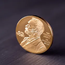
Image of Nobel Prize medal on a table.

Who Will Win the 2026 Nobel Prize? Scientists Make Their Predictions

A photograph of a rattlesnake against a jungle-like background.

Vipers’ Self-Defense Mechanism Could Spark a New Antivenom Strategy

Sex-Specific Dioxin Toxicity in a Humanized AHR Model

Sex-Specific Dioxin Toxicity in a Humanized AHR Model

Miltenyi
Alt-Text A conceptual image of blue histograms on a graph gradually increasing in size up toward the right with a trendline following.

Federal Research Grant Awards Fall to a Six-Year Low

Multimedia

Evaluating Blood Biomarkers for Alzheimer’s Disease Staging

Evaluating Blood Biomarkers for Alzheimer’s Disease Staging

​Beckman Coulter
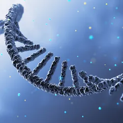
Three-dimensional illustration of a curved, single-stranded RNA molecule against a blue background

Gene Editing Precision Depends on Guide RNA Quality

TriLink BioTechnologies
The Scientist October Digest cover
October 2026

Antarctica’s Blood Falls Teems With Mysterious Microbial Life

Miles away from the closest ocean, marine microbes thrive in the red ice and mud around Blood Falls, offering clues to microbial adaptation and past climate events.

View this Issue
Evaluating Blood Biomarkers for Alzheimer’s Disease Staging

Evaluating Blood Biomarkers for Alzheimer’s Disease Staging

​Beckman Coulter
Find better biologics faster with Stunner and the AI Developability Workcell

Find better biologics faster with Stunner and the AI Developability Workcell

Unchained Labs
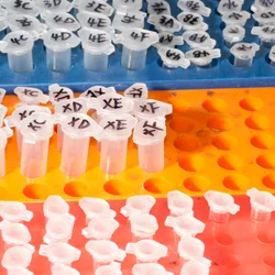
An image of microcentrifuge tubes arranged within colorful laboratory racks.

Choose clarity. Choose speed. Choose MyMass™

Refeyn
Characterizing LNP Heterogeneity With Super-Resolution Imaging

Characterizing LNP Heterogeneity With Super-Resolution Imaging

Oni

Products

Stellaromics Logo

Stellaromics Expands Pyxa Adoption as Instruments are Installed at Leading Research Institutions Across the U.S. and EuropeStellaromics, Inc., the leader in 3D

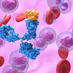
A monoclonal antibody surrounded by immune cells and red blood cells

CellRaft AIR: Find the Right Antibody-Producing Cells Faster

Cell Microsystems
Maxwell® Rapid ccfDNA Kit

Maxwell® Rapid ccfDNA Kit: Automated Purification of ccfDNA in Under 30 Minutes

Promega
Leica Biosystems, Tempus, and Paige logos side by side, marking the addition of Paige digital pathology AI tools to the Aperio AI Store.

Oncology Researchers Gain AI Cancer Tools in One Platform