SKIP ODONNELL/ISTOCKPHOTO.COM

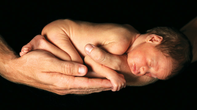
Louisa Corinne Grant just couldn’t wait to make her grand entrance into the world. My second child and first daughter was born on January 3, 2013, at 8:21 p.m., surprising me and my wife Kerry by arriving at 35 weeks of gestation, about a month before her expected due date.

Kerry had experienced an exceedingly normal pregnancy and exhibited none of the warning signs or risk factors for premature birth. At some point during her pregnancy, however, a complicated cascade of signaling and chemical crosstalk short-circuited. Something among the gene- and protein-driven pathways designed to keep Louisa wrapped in the warmth of her mother’s womb for a full 40 weeks went haywire. And the molecular confusion caused baby Louisa to depart from in utero comfort before she was fully prepared for the rigors of the outside world.

As Kerry and I watched our daughter navigate (with the...

We were surprised to learn that the conditions that trigger preterm birth, which is defined as any birth taking place earlier than 37 weeks of gestation, remain virtually unknown to science even though it affects 1 out of every 9 babies born in the United States, according to the Centers for Disease Control and Prevention (CDC). Knowledge of the molecular triggers for normal-term birth has proven almost equally elusive. But researchers around the world are seeking answers to these vexing questions, and, in the process, developing a fundamental understanding of human gestation and labor that could help save thousands of young lives every day.

It is a surprise that a phenomenon such as labor is essential for the survival of our species, and yet we know very little about the mechanisms that control it.—­ Roberto Romero, Wayne State Uni­versity

“It is a surprise that a phenomenon such as labor is essential for the survival of our species, and yet we know very little about the mechanisms that control it,” says Roberto Romero, Wayne State University perinatologist and head of the Program for Perinatal Research and Obstetrics at the National Institutes of Health’s National Institute of Child Health and Human Development. “We need a molecular taxonomy of premature labor,” he adds. “This is an intellectual challenge that is unlike many others that have been posed by biology and medicine.”

Strange bedfellows

From a physiological perspective, pregnancy and labor are exercises in extremes. After one sperm among hundreds of millions fertilizes an egg (itself a pretty remarkable event), the resulting embryo implants in the lining of the uterus, and the developing fetus—essentially a foreign entity—takes up residence inside the body of a host whose natural immunologic impulse would be to expel an invader as quickly as possible.

But instead of outright rejection and expulsion, a mysterious chemical conversation between mother and child ensues. Through a tangle of signaling pathways that researchers are just now beginning to tease apart, mother—her immune system modulated—ensconces baby in the serenity of a uterus that expands to 500 times its normal size, providing a retreat that allows development to progress for an average of 40 weeks in humans. But even more puzzling than the intricacies of the immune system modulation that allows mother to tolerate a pregnancy for the better part of a year is the cascade of events at the end of that period, when the process of birth, or parturition, commences.

When the time for parturition arrives, a message to contract is delivered to the smooth muscles in the wall of the uterus, a layer called the myometrium. In doing so, the organ that has stretched to such an amazing capacity becomes, for a brief time, one of the strongest muscles in the human body (by weight), exerting an incredible force downward and outward upon the infant, hell-bent on expelling it and the placenta from the body through the narrow birth canal.

The intricate steps of this process in humans are essentially mysterious to science, perhaps more so than any other physiological phenomenon our species hosts. Some of the players—hormones like progesterone and a few of its receptors—are known, but the sequence of molecular events, and many members of its cast of chemicals and cells, are as yet unidentified.

But that is beginning to change.

Prior thinking

The insight that is propelling the field of reproductive science forward in its quest to understand the molecular roots of pregnancy and labor is a growing appreciation of the complexity of the phenomena. Much as with cancer, the terms “preterm labor” and “preterm birth” convey a simplicity that does not exist. “Preterm birth is an oddity in that it is one of the few disorders that’s defined by a calendar event rather than some pathogenesis,” says Stephen Lye, vice chair of research and professor of obstetrics and gynecology at the University of Toronto. “We’re now understanding that preterm birth is probably a whole series of different diseases. That’s exciting in that it now puts us in the position to say, ‘I’m going to focus on a fairly defined phenotype.’ ”

Previously, researchers and clinicians sought to understand normal labor and birth, and therefore addressed the problem of preterm labor and birth at the level of the uterus. The key event in labor is the contraction of that muscular organ. If uterine contractions—the rhythmic waves that represent pregnancy’s endgame—could be stopped, the reasoning went, birth could be delayed temporarily.

A class of drugs called tocolytics, which are still used today, can indeed delay a preterm birth for up to a few days by slowing or stopping contractions of the uterus that might otherwise expel a dangerously premature baby. This can buy precious time for other interventions, such as the administration of glucocorticoid drugs that can speed fetal lung development. But tocolytics come with their own suite of risks and cannot be used in many scenarios. And from a conceptual standpoint, uterine contractions—premature or otherwise—come at the end of a series of events that precede the main event of labor by weeks. If researchers can look further upstream into the molecular cascades that signal the end of pregnancy and the start of parturition, there is the potential to identify a trigger that can be targeted long before the initiation of contractions.

Out of necessity, nearly all the labs seeking to map the upstream dynamics that ultimately lead to contractions and birth root themselves in one of the few clear-cut realities regarding human gestation: the steroid hormone progesterone plays a key role in maintaining pregnancy. At the start of pregnancy and during the fertilized egg’s journey to the uterus for implantation, progesterone is produced by the corpus luteum, a temporary mass of tissue derived from the ovarian follicle that expelled the egg before fertilization. Later, the placenta pumps out progesterone, keeping systemic levels in the mother high throughout pregnancy, which decreases the contractility of uterine muscle. Seminal experiments in the 1930s and ’40s by University of Rochester Medical School anatomist George Corner—the codiscoverer of progesterone—showed that without the steroid hormone women could not become pregnant, nor could a pregnancy be maintained.

Though the link between progesterone and the maintenance of pregnancy has been thoroughly established, very little is known about how exactly the hormone works when the time comes for gestation to end. There are likely dozens of mechanisms that act to set off labor at the end of pregnancy, says Sam Mesiano, a Case Western Reserve University physiologist, but only one—with progesterone at its center—to block it. “The key is to find where all the pathways converge and attack the convergent point therapeutically as the target,” he says. And that is exactly what a bevy of research teams is seeking to do, generating some interesting models regarding the roles played by genes, the immune system, and the body’s inflammatory machinery.

O'LUK/ISTOCKPHOTO.COM

Uniquely Human

Animal models are essential for gaining an appreciation of human physiology. But mice and fruit flies have not been as helpful in understanding human pregnancy and parturition as they have been in elucidating other physiological phenomena. The reason: when nonhuman animals procreate, gestation and birth seem to proceed in a much more predictable fashion than in humans. In sheep, for example, decades of experiments have shown that fetal production of the adrenal hormone cortisol, directed by its pituitary gland, initiates labor. In mice, on the other hand, the main signal to commence labor emanates from the mother’s tissues. But in humans, researchers are still seeking to understand the contributions of a complex tangle of pathways, both fetal and maternal that signal the maintenance of pregnancy and the initiation of labor.

Human pregnancy and birth are distinct from all the varied reproductive realities of our animal kin because evolution set humans on a novel trajectory, not just mentally and socially, but also, on a more basic level, physiologically. Upright walking and narrow pelvises conferred distinct advantages upon our ancestors as they roamed African savannahs. Additionally, natural selection gave them bigger brains, which necessitated encephalization, or increasing skull size. But both of these adaptations require giving birth to less-developed young: big-headed babies need to exit a narrower birth canal, the so-called “obstetrical dilemma.” This, argues Sam Mesiano of Case Western Reserve University in Ohio, may have perturbed the coordinated pathways that control the timing of birth in other mammals. “We had a unique evolutionary process that selected for our timing mechanism,” he says. “The benefits of encephalization outweighed the cost of selecting a timing mechanism that was really sloppy.”

Other mammals deliver their babies on much stricter schedules than humans. According to Leonardo Pereira, a perinatologist at Oregon Health & Science University, pregnant mice will all deliver their offspring on day 20 or 21 of gestation. Monkeys will deliver on day 181 or 182, without much variation. Humans, on the other hand, “deliver in a 6-week range,” Pereira says. “The system is not very tightly regulated.”

Not only do humans have what appears to be a vastly more complicated system for signaling the timing of birth, but human babies born at term are underdeveloped in comparison to those of our closest primate relatives. Chimpanzee babies, for example, can grasp their mother’s fur (a skill essential to their survival) immediately after being born. Human newborns, while they still retain a vestigial grasp reflex that functions immediately after birth, would be hopeless at holding on to a mother gamboling about in the treetops. “What we think happened evolutionarily is that the timing mechanism for human birth was changed so that babies came out earlier than they should,” Mesiano says.

This disparity between humans and other animals has led to a historical reluctance to use animal models to understand human pregnancy and birth. But that attitude is changing. “We didn’t have an appropriate animal model,” says University of Pittsburgh cell biologist Jennifer Condon-Jeyasuria, who works with mouse models to elucidate the molecular mechanics of pregnancy and labor. “That’s changed. Even though the mouse does have some limitations, most of the field would be quite happy modeling in the mouse.”

As researchers unravel the snarl of pathways that play a role in both maintaining pregnancy and initiating labor, animal models will prove vital. Already, rhesus macaques have offered key insights into intrauterine infection, which is thought to cause about half of all preterm human births. And mice are yielding important information on the genetic and immune modulations and inflammatory pathways that seem to contribute to human gestation and labor.

Plausible models

Carole Mendelson, a reproductive biologist at the University of Texas Southwestern Medical Center, has made great strides in elucidating the molecular and cellular machinery behind progesterone’s effects on pregnancy. The conceptual starting point for her work is the fact that in virtually every nonhuman mammalian species, such as the mice she uses as model organisms, levels of progesterone decline dramatically at the end of pregnancy. In humans, however, circulating progesterone levels do not drop off. To Mendelson and many of her peers, this means that for human labor to begin there must be changes involving progesterone receptors within myometrial cells that make the receptors less responsive to the flood of progesterone, a phenomenon called “functional progesterone withdrawal.”

“There have to be things taking place to impair the function of the progesterone receptor near term,” Mendelson says.

Her work centers on interactions between a family of microRNAs (miRNAs), miR-200, and the target transcription factors, called zinc finger E-box binding homeobox proteins, ZEB1 and ZEB2. First using gene expression arrays and more recently with human tissues in vitro and mouse models of preterm labor, Mendelson and her colleagues have shown that miR-200 miRNAs, ZEB1, and ZEB2 act as genetic switches in uterine tissues during the transition from pregnancy to labor.1

Here’s how it works: during pregnancy, when progesterone binds to progesterone receptors, myometrial cells begin producing more ZEB1, which inhibits the expression of genes coding for contractile proteins essential for the uterine contractions that define labor. ZEB1 and ZEB2 also inhibit the expression of the miR-200 family, and their decline further upregulates the ZEBs. This feedback loop maintains the uterus in a quiescent, noncontractile state for the full duration of pregnancy. But when progesterone receptor function becomes altered, levels of the ZEBs drop, contractile proteins are expressed, and miR-200 levels rise rapidly. This cascade ultimately leads to the wholesale uterine contractions of labor. (See illustration below.)

Cell biologist Jennifer Condon-Jeyasuria, of the University of Pittsburgh, has proposed additional molecular mechanisms that could underlie progesterone’s maintenance of uterine quiescence during pregnancy. She and colleagues have shown that levels of caspase 3, an enzyme linked to reduced contractile ability in a variety of muscle cell types, are kept high during pregnancy through the action of progesterone, and that this contributes to the degradation of uterine contractile proteins in mice. Prior to the onset of labor, when functional progesterone withdrawal occurs, caspase 3 levels drop and myometrial contractile proteins are left to do their work.2

Some researchers have shifted their focus further upstream from the miR200 miRNAs, their target ZEBs, and caspase 3 to study progesterone receptors and the mechanisms behind their ability to cause functional progesterone withdrawal, which may then set off downstream signaling cascades that initiate labor. Lye and colleagues, for example, recently proposed a model in which high levels of a coregulator of the progesterone receptor could play a crucial role in repressing the expression of contractile proteins, such as connexin 43 (Cx43), during pregnancy. As the end of gestation approaches, levels of the coregulator, polypyrimidine tract-binding protein-associated splicing factor (PSF), drop in response to mounting mechanical stretch signals from the myometrium and a rise in estrogen levels. This loosens the transcriptional control that the progesterone receptor had been exerting over genes that express contractile proteins, and ushers in the start of labor.3 (See illustration below.)

Immune system involvement

Lye has also studied the immune cascade set in motion by the stretching of myometrial cells as the fetus increases in size. He and his collaborators have suggested that uterine stretching signals the cells of the myometrium to secrete increasing amounts of pro-inflammatory cytokines (including IL-6 and IL-9) and chemokines (including CCL-2, CCL-7, and others), which activate white blood cells associated with inflammation, such as neutrophils and macrophages, further increasing the production of cytokines and chemokines and exacerbating inflammation.4 (See illustration below.) This cascade could aid in triggering the onset of labor by ramping up the expression of contractile-associated proteins, including receptors for hormones that serve to remodel uterine and cervical tissue to ready the mother’s body for parturition.

Furthermore, Lye and his collaborators have begun to describe a proinflammatory cytokine signature that could be used to predict the onset of both preterm and term labor.5 “We’ve thought about a model in which towards the end of pregnancy there are changes that go on within the muscle layer and the lining of the uterus that produce and release cytokines into the mother’s circulation, and subsets of those cytokines seem to be able to activate subsets of the immune cells and target the uterus,” Lye says. “Hormone pathways were important for the onset of labor, but they weren’t enough by themselves. Our thinking is you can find inflammation in almost all labor situations.”

MOLECULAR MODELS OF BIRTH
View full size JPG | PDF
LUCY READING-IKKANDA
Previously, Condon-Jeyasuria, while doing a postdoc in Mendelson’s lab, showed that a chemical produced by the maturing fetal lung could play a role in initiating labor. She and her colleagues detected a major lung-surfactant protein, surfactant protein A (SP-A), in the amniotic fluid of pregnant mice. SP-A levels rose throughout pregnancy and drove concomitant rises in cytokine expression by macrophages in the amniotic fluid as well as the activation of the NF-κB protein complex, a transcription factor that modulates immune responses. The researchers could induce preterm labor by injecting additional SP-A into the amniotic fluid earlier in pregnancy.6 (See illustration, right.)

Inflammation is likely especially important in preterm labor, as infection, which typically sets off an inflammatory response, is implicated in a significant proportion of preterm births. According to Romero, 20 percent of women experiencing preterm birth test positive for microorganisms in the amniotic fluid that surrounds the fetus in the uterus.

Using rhesus macaque models of preterm birth, physiologist Peta Grigsby, head of the Pregnancy and Perinatal Research Group in the Division of Reproductive Sciences at the Oregon National Primate Research Center, has produced convincing data that indicate the potential benefit of treating amniotic infection with antibiotics in order to prolong pregnancy. She and her collaborators have surveyed the molecular dynamics that result from introducing Ureaplasma, a common bacterium that inhabits both male and female genitalia and has been implicated in preterm birth, into the amniotic fluid surrounding macaque fetuses. By taking a longitudinal look at cytokines, prostaglandins, and other markers, such as hypoxia in the fetus and maternal white blood cell counts, Grigsby and her colleagues have shown that both mother and baby mount immune and inflammatory responses to the infection. They’ve shown that they can delay labor for about 10 days in an infected mother monkey by administering intravenous antibiotics, but can’t altogether stop the animal from delivering early.7 “Antibiotics are not going to stop the prostaglandins or the cytokines,” Grigsby says. “Once that chain of events has started, it’s very difficult to stop if you’re just targeting the bacteria.”

Other researchers are seeking to add more detail to the emerging picture of the molecular milieu that attends pregnancy and birth. Ongoing studies are striving to characterize the vaginal and uterine microbiomes’ dynamic effect on pregnancy and labor8 and to identify the full complement of genes that are involved in the processes.9

Though the lack of clarity concerning the mechanics of pregnancy and parturition can be disconcerting, the beauty of studying their molecular intricacies is that the field is so wide open. “There are probably thousands of default pathways to allow this basic fundamental process to occur,” says Condon-Jeyasuria.

And bringing modern experimental tools to the quest, especially those used to probe immune dynamics, is changing the game, according to Sing Sing Way, a Cincinnati Children’s Hospital infectious disease researcher who studies the lasting effects of pregnancy on the mother’s immune system. “Only now are what I consider the best immunological tools being applied to this problem of pregnancy and preterm labor,” says Way, who has recently published research showing that pregnancy selectively stimulates the accumulation of maternal FOXP3+ CD4 cells, allowing for the persistence of maternal regulatory T cells that expand more rapidly with a second pregnancy.10 “Up until now, the people who are studying this problem haven’t been immunologists.” This immunological phenomenon, like so many of the other steps on the path to labor, may ultimately be controlled by progesterone, Way adds. “There’s a direct association between progesterone levels in pregnancy and the expansion of fetal-specific regulatory T cells,” he says. “Whether that’s a cause and effect is what we’re trying to prove now.”

The impact

Though basic research has yet to fully map out the complexities of human gestation and birth, epidemiology has shown that embedded within those phenomena is a propensity for mishap. The 12 percent of US babies every year who are born preterm—my daughter among them—add up to more than half a million infants.

Global statistics tell a still grimmer tale of the perils of being born early. According to the World Health Organization, 15 million babies around the world are born early every year, most in Africa and South Asia. And about 1 million of these babies die from complications associated with their truncated gestation, making preterm birth the leading killer of newborns the world over. Overall, babies born preterm die 120 times more frequently than babies born at term.

Even though she was denied the full term of her gestation, Louisa Grant was lucky. After her unexpectedly early entry into the world, she required only a couple of days of artificial-light treatment to ramp up the elimination of bilirubin, a waste product of the breakdown of red blood cells. Louisa was what is called “late preterm,” which means she was born between 32 and 37 weeks of gestation. Premature babies can also be “extremely preterm,” born after less than 28 weeks of gestation, or “very preterm,” which means they were born between 28 and 32 weeks, according to the World Health Organization. Since her birth, Louisa has thrived, gaining weight and developing normally.

But for babies that survive the ordeal of being born even earlier than Louisa, a gauntlet of health and developmental problems awaits. After weeks or months spent in a neonatal intensive care unit—hooked up to ventilators, connected to intravenous drips, intubated to ensure adequate nutrition—babies born extremely or very preterm can be plagued with intellectual disabilities, cerebral palsy, vision problems, hearing deficits, and feeding and digestive difficulties. Altogether, preterm birth costs the US health-care system more than $26 billion every year, according to the CDC.

Until we actually look at this in a much more sophisticated way, we’re not going to solve the human problem.—­ David Stevenson,
Stanford University School of Medicine

There has, however, been recent progress in reversing this bleak trend. Even though the rate of premature birth has risen steadily in the United States over the past 30 years or so, it has inched downward in the past several years—from 12.8 percent in 2006 to 11.7 percent in 2011. Much of this success likely lies in the current clinical practice of administering the hormone progesterone to pregnant women who had a prior preterm birth.

But because the detailed mechanics of this progesterone treatment are still unknown, it remains a blunt weapon in the fight to prolong pregnancy and reduce the risk of premature birth. The real key to preventing preterm labor and birth, according to David Stevenson, a Stanford University School of Medicine neonatologist and director of a new March of Dimes–funded Prematurity Research Center, involves understanding the molecular roots of labor. “A lot of the work that we are trying to do is to literally understand the pathogenesis of preterm birth in a more fundamental way,” he says. “Nothing has worked for over 30 or 40 years. Until we actually look at this in a much more sophisticated way, we’re not going to solve the human problem.” 

References

  1. N.E. Renthal et al., “miR-200 family and targets, ZEB1 and ZEB2, modulate uterine quiescence and contractility during pregnancy and labor,” PNAS, 107:20828-33, 2010.
  2. P. Jeyasuria et al., “Progesterone-regulated caspase 3 action in the mouse may play a role in uterine quiescence during pregnancy through fragmentation of uterine myocyte contractile proteins,” Biol Repro, 80:928-34, 2009.
  3. N. Xie et al., “Expression and function of myometrial PSF suggest a role in progesterone withdrawal and the initiation of labor,” Mol Endocrinol, 26:1370-79, 2012.
  4. O. Shynlova et al., “Physiologic uterine inflammation and labor onset: integration of endocrine and mechanical signals,” Repro Sci, 20:154-67, 2013.
  5. O. Shynlova et al., “Cytokine signature associated with the onset of term and preterm labour,” Reprod Sci, 183(suppl):151A, 2011.
  6. J.C. Condon et al., “Surfactant protein secreted by the maturing mouse fetal lung acts as a hormone that signals the initiation of parturition,” PNAS, 101:4978-83, 2004.
  7. P.L. Grigsby et al., “Maternal azithromycin therapy for Ureaplasma intraamniotic infection delays preterm delivery and reduces fetal lung injury in a primate model,” Am J Obstet Gynecol, 207:475.e1-14, 2012.
  8. R.M. Brotman et al., “Association between Trichomonas vaginalis and vaginal bacterial community composition among reproductive-age women,” Sex Transm Dis, 39:807-12, 2012.
  9. S.M. Dolan, I. Christiaens, “Genome-wide association studies in preterm birth: implications for the practicing obstetrician-gynaecologist,” BMC Pregnancy and Childbirth, 13(Suppl 1):S4, 2013.
  10. 10. J.H. Rowe et al., “Pregnancy imprints regulatory memory that sustains anergy to fetal antigen,” Nature, 490:102-06, 2012.

Interested in reading more?

Magaizne Cover

Become a Member of

Receive full access to digital editions of The Scientist, as well as TS Digest, feature stories, more than 35 years of archives, and much more!
Already a member?