3-D X-Rays Catch Insects in Flight

Using a new approach, researchers capture the in vivo mechanics of a flying fly.

Written byKerry Grens
| 2 min read

Register for free to listen to this article
Listen with Speechify
0:00
2:00
Share

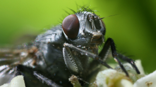
WIKIMEDIA, MAKRO FREAKScientists have developed a technique to see inside the muscles of a blowfly in flight. The flies were tethered, but moving their wings, as X-ray microtomatography captured the internal mechanics of how their steering muscles operate. “This by itself is a grand achievement at a wingbeat frequency of 145 Hz,” wrote Anders Hedenström of Lund University in Sweden, in a commentary in PLOS Biology.

Videos of the flies, published in PLOS Biology this week (March 25), offer an unprecedented look at the inner workings of the flight muscles as a tethered fly is spun in a circle. “This has been an awe-inspiring project on so many levels, not least the exquisite complexity of the insects themselves, but seeing the 3-D movies render for the first time was one of those breakthrough moments that as a scientist I’ll never forget,” Graham Taylor of Oxford University, who led the work, told PLOS Biologue.

Hedenström pointed out that, while the study represents a “methodological breakthrough,” a tethered fly is not the same as a freely moving fly, which could roll, speed up, and stop. Also, ...

Interested in reading more?

Become a Member of

The Scientist Logo
Receive full access to more than 35 years of archives, as well as TS Digest, digital editions of The Scientist, feature stories, and much more!
Already a member? Login Here

Related Topics

Meet the Author

  • kerry grens

    Kerry served as The Scientist’s news director until 2021. Before joining The Scientist in 2013, she was a stringer for Reuters Health, the senior health and science reporter at WHYY in Philadelphia, and the health and science reporter at New Hampshire Public Radio. Kerry got her start in journalism as a AAAS Mass Media fellow at KUNC in Colorado. She has a master’s in biological sciences from Stanford University and a biology degree from Loyola University Chicago.

    View Full Profile
Share
Illustration of a developing fetus surrounded by a clear fluid with a subtle yellow tinge, representing amniotic fluid.
January 2026, Issue 1

What Is the Amniotic Fluid Composed of?

The liquid world of fetal development provides a rich source of nutrition and protection tailored to meet the needs of the growing fetus.

View this Issue
Skip the Wait for Protein Stability Data with Aunty

Skip the Wait for Protein Stability Data with Aunty

Unchained Labs
Graphic of three DNA helices in various colors

An Automated DNA-to-Data Framework for Production-Scale Sequencing

illumina
Exploring Cellular Organization with Spatial Proteomics

Exploring Cellular Organization with Spatial Proteomics

Abstract illustration of spheres with multiple layers, representing endoderm, ectoderm, and mesoderm derived organoids

Organoid Origins and How to Grow Them

Thermo Fisher Logo

Products

Brandtech Logo

BRANDTECH Scientific Introduces the Transferpette® pro Micropipette: A New Twist on Comfort and Control

Biotium Logo

Biotium Launches GlycoLiner™ Cell Surface Glycoprotein Labeling Kits for Rapid and Selective Cell Surface Imaging

Colorful abstract spiral dot pattern on a black background

Thermo Scientific X and S Series General Purpose Centrifuges

Thermo Fisher Logo
Abstract background with red and blue laser lights

VANTAstar Flexible microplate reader with simplified workflows

BMG LABTECH