Eye evolution questioned

Invertebrates with vertebrate-like vision challenge the idea that the two groups of organisms have distinctly different visual receptors

Written byAmy Maxmen
| 2 min read

Register for free to listen to this article
Listen with Speechify
0:00
2:00
Share
The standing dogma of eye evolution is challenged with the discovery of an invertebrate that sees light like vertebrates do, rather than like their more closely related cousins, according to a study published today (March 1) in EvoDevo.
T. transversa larva
Image: Nina Furchheim, Berlin Museum of Natural History
"Now the story is more complicated than it was before, when we thought there was a clear-cut division between vertebrates and invertebrates," said lead author linkurl:Yale Passamaneck;http://www.kewalo.hawaii.edu/martindale/mmpeople.html from Kewalo Marine Laboratory at the University of Hawaii.Animal eyes vary in appearance, but the light-sensing photoreceptor cells within them come in just two varieties: ciliary and rhabdomeric. Vertebrates see light with the ciliary type, which sports a folded, hair-like cilium, while invertebrates see with rhabdomeric photoreceptors, which typically bear bristles. In 2004, biologists hypothesized that an ancestor of both invertebrates and vertebrates sensed light with rhabdomeric receptors -- but also had ciliary receptors embedded deep within their brains, where they have been found in marine worms and bees, and might have sensed patterns, such as the lunar cycle. As vertebrates evolved, ciliary receptors theoretically migrated towards the body surface and became the primary tools for sight. But the new results challenge this scenario with the discovery of surface ciliary photoreceptors in the larvae of marine invertebrates called brachiopods (Terebratalia transversa), shelled creatures that have been around for 540 million years. Passamaneck and his colleagues identified the expression of a gene characteristic of ciliary receptors, ciliary-opsin, in cells in the light-sensing "eyespot" of larval brachiopods. "This is completely novel," said linkurl:Detlev Arendt,;http://www.embl.de/research/units/dev_biology/arendt/members/?s_personId=673 an evolutionary biologist at the European Molecular Biology Laboratory in Germany who was not involved with the study. "No one has looked for opsins in many animals, and this is exactly what we should be doing."
T. transversa adult
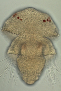
Image: Yale Passamaneck
Now it's unclear which photoreceptor originally gave animals sight, and which kind evolved to sense light later. Or, perhaps an ancestor used both receptors to see, and over the millennia, one variety or the other lost its visual function. Additionally, the researchers found that sand-grain-sized brachiopod embryos, which don't have a head, nerves, or eyespots, also expressed the ciliary-opsin gene, suggesting it may have a role even before the development of true eyes with photoreceptors that are connected to the brain. Found in nearly half of the embryos' cells, this is the first report of ciliary-opsin expression in non-neuronal cells. "We're really excited about this discovery because it was really a naïve experiment to do since these embryos don't even have eyes," Passamaneck says. While the function of ciliary-opsin at this life stage remains unclear, the embryos did gravitate towards light, hinting at the possibility of a role in rudimentary vision. Playfully dubbed "swimming eyeballs" by the team, the embryos may represent a time before true eyes evolved, when visual proteins were somehow involved with sensing light in individual cells, Passamaneck said. If so, brachiopods may provide key insights into how vision first evolved. Y. Passamaneck, et al., "Ciliary photoreceptors in the cerebral eyes of a protostome larva," linkurl:EvoDevo,;http://www.evodevojournal.com/content/2/1/6/abstract 2:6, 2011.
**__Related stories:__***linkurl:Seeing ear to eye;http://www.the-scientist.com/blog/display/57540/
[1st July 2010]*linkurl:Ancient eyes head for the light;http://www.the-scientist.com/blog/display/55204/
[19th November 2008]*linkurl:An eye on history;http://www.the-scientist.com/news/display/54992/
[29th August 2008]
Interested in reading more?

Become a Member of

The Scientist Logo
Receive full access to more than 35 years of archives, as well as TS Digest, digital editions of The Scientist, feature stories, and much more!
Already a member? Login Here

Meet the Author

Share
December digest cover image of a wooden sculpture comprised of multiple wooden neurons that form a seahorse.
December 2025, Issue 1

Wooden Neurons: An Artistic Vision of the Brain

A neurobiologist, who loves the morphology of cells, turns these shapes into works of art made from wood.

View this Issue
Stacks of cell culture dishes, plates, and flasks with pink cell culture medium on a white background.

Driving Innovation with Cell Culture Essentials

Merck
Stacks of cell culture dishes, plates, and flasks with pink cell culture medium on a white background.

Driving Innovation with Cell Culture Essentials

MilliporeSigma purple logo
Human iPSC-derived Models for Brain Disease Research

Human iPSC-derived Models for Neurodegenerative Disease Research

Fujifilm
Abstract wireframe sphere with colorful dots and connecting lines representing the complex cellular and molecular interactions within the tumor microenvironment.

Exploring the Inflammatory Tumor Microenvironment 

Cellecta logo

Products

brandtech logo

BRANDTECH® Scientific Announces Strategic Partnership with Copia Scientific to Strengthen Sales and Service of the BRAND® Liquid Handling Station (LHS) 

Top Innovations 2026 Contest Image

Enter Our 2026 Top Innovations Contest

Biotium Logo

Biotium Expands Tyramide Signal Amplification Portfolio with Brighter and More Stable Dyes for Enhanced Spatial Imaging

Labvantage Logo

LabVantage Solutions Awarded $22.3 Million U.S Customs and Border Protection Contract to Deliver Next-Generation Forensic LIMS