 |
cell membrane Image: Charles Lieber |
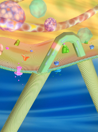 |
Image: Charles Lieber |
**__Related stories:__***linkurl:Single neuron power;http://www.the-scientist.com/blog/display/55091/
[15th October 2008]*linkurl:The brain in silico;http://www.the-scientist.com/blog/display/53839/
[5th November 2007]*linkurl:Monitoring Neural Activity In Vivo;http://www.the-scientist.com/article/display/14959/
[27th September 2004]


