Ancient eyes head for the light

Simple communication between a pair of neighboring cells allows tiny marine worms to move toward light using a sensory organ believed to be an ancient precursor of the eye, according to a study out this week in Nature. "It's remarkable that a primitive organism of the ocean, a living marine zooplankton, has the sophisticated ability to move in response to light with a pigment-photoreceptor cell combination," linkurl:Russell Fernald,;http://www.stanford.edu/group/fernaldlab/index.shtml an evolu

Written byJennifer Evans
| 2 min read

Register for free to listen to this article
Listen with Speechify
0:00
2:00
Share
Simple communication between a pair of neighboring cells allows tiny marine worms to move toward light using a sensory organ believed to be an ancient precursor of the eye, according to a study out this week in Nature. "It's remarkable that a primitive organism of the ocean, a living marine zooplankton, has the sophisticated ability to move in response to light with a pigment-photoreceptor cell combination," linkurl:Russell Fernald,;http://www.stanford.edu/group/fernaldlab/index.shtml an evolutionary neuroethologist at Stanford who was not involved in the study, told The Scientist. He added that the paper provides "proof that a simple pair of cells can give directive information." The vertical migration of plankton in response to light constitutes the largest biomass transport on Earth. Researchers have believed that the eyespots of multicellular zooplankton larvae, comprised of a single photoreceptor and shading pigment cell, are responsible for phototaxis, or movement toward light, but the mechanism has never been identified. Eyespots allow organisms to sense the direction of light but not make out images. linkurl:Detlav Arendt,;http://www-db.embl.de/jss/EmblGroupsOrg/per_673.html a molecular biologist at the European Molecular Biology Laboratory in Germany and senior author on the Nature study, spent the past 10 years studying eyespots in the marine rag-worm Platynereis. The rag-worm is covered in cilia, which it uses to propel itself forward in a helical fashion towards light.

The eyespots of Platynereis larvae allow it to move toward light

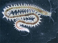
In linkurl:2001,;http://www.ncbi.nlm.nih.gov/pubmed/11604122?ordinalpos=3&itool=EntrezSystem2.PEntrez.Pubmed.Pubmed_ResultsPanel.Pubmed_DefaultReportPanel.Pubmed_RVDocSum Arendt's group discovered that the gene linkurl:Pax6,;http://www.the-scientist.com/article/display/19190/ which controls the development of eyes and other sensory organs in humans, was also active in rag-worm eyespots, confirming early traces of more sophisticated eye systems. In linkurl:2004,;http://www.ncbi.nlm.nih.gov/pubmed/15514158?ordinalpos=&itool=EntrezSystem2.PEntrez.Pubmed.Pubmed_ResultsPanel.SmartSearch&log$=citationsensor they identified two types of photoreceptor cells in the rag-worm -- the rhabdomeric photoreceptors in the eyespots and ciliary photoreceptors in the brain. In their latest set of experiments, the researchers used linkurl:immunohistochemistry;http://www.the-scientist.com/article/display/17060/ to trace the photoreceptor axons directly to neighboring ciliated cells in Platynereis larvae. "Essentially there is no nervous system in between" the photoreceptors and ciliated cells, Arendt said. "The axons contact ciliated cells in the neighborhood." Double whole-mount in situ hybridization revealed that photoreceptors and ciliated cells were communicating via the neurotransmitter acetylcholine. Blocking the acetylcholine receptor prevented the larvae's helical movement toward light without affecting the speed at which they swam, revealing the direct sensory-motor coupling between the photoreceptor and the cilia cells. To understand how light exposure influences the movement of cilia, they selectively illuminated one of the organism's two eyespots and recorded the force of cilia beats. The light activated five ciliated cells neighboring the eyespot, which resulted in a slowed beat with more force on one side of the larvae without affecting the cilia on the opposing side of the larvae, leading the larvae to make a helical turn. linkurl:John Spudich,;http://www.uth.tmc.edu/bmb/faculty/Spudich/Spudich.html a photobiologist at the University of Texas in Houston who was not involved in the study, told The Scientist that Arendt's "elegant study" identifies a photosensory mechanism that may be the "key evolutionary intermediate suggested by Charles Darwin's reflections on the evolution of human vision." Previous studies in Spudich's lab showed the unicellular algae Chlamydomonas also controls helical phototactic swimming by modulating asymmetric ciliary beating. The study also has ecological implications, Arendt said. "Phototaxis is of important component of marine life," he said. "If you want to understand anything in the ecosystem, including planet climate change, you need to start by understanding the mechanism by which [phototaxis is taking place]." Image courtesy of Arendt, D. and Jekely, G., EMBLThis post has been updated from a previous post to correctly identify Russell Fernald of Stanford University. The Scientist regrets the error.
Interested in reading more?

Become a Member of

The Scientist Logo
Receive full access to more than 35 years of archives, as well as TS Digest, digital editions of The Scientist, feature stories, and much more!
Already a member? Login Here

Meet the Author

Share
December digest cover image of a wooden sculpture comprised of multiple wooden neurons that form a seahorse.
December 2025, Issue 1

Wooden Neurons: An Artistic Vision of the Brain

A neurobiologist, who loves the morphology of cells, turns these shapes into works of art made from wood.

View this Issue
Stacks of cell culture dishes, plates, and flasks with pink cell culture medium on a white background.

Driving Innovation with Cell Culture Essentials

Merck
Stacks of cell culture dishes, plates, and flasks with pink cell culture medium on a white background.

Driving Innovation with Cell Culture Essentials

MilliporeSigma purple logo
Abstract wireframe sphere with colorful dots and connecting lines representing the complex cellular and molecular interactions within the tumor microenvironment.

Exploring the Inflammatory Tumor Microenvironment 

Cellecta logo
An image of a DNA sequencing spectrum with a radial blur filter applied.

A Comprehensive Guide to Next-Generation Sequencing

Integra Logo

Products

brandtech logo

BRANDTECH® Scientific Announces Strategic Partnership with Copia Scientific to Strengthen Sales and Service of the BRAND® Liquid Handling Station (LHS) 

Top Innovations 2026 Contest Image

Enter Our 2026 Top Innovations Contest

Biotium Logo

Biotium Expands Tyramide Signal Amplification Portfolio with Brighter and More Stable Dyes for Enhanced Spatial Imaging

Labvantage Logo

LabVantage Solutions Awarded $22.3 Million U.S Customs and Border Protection Contract to Deliver Next-Generation Forensic LIMS