
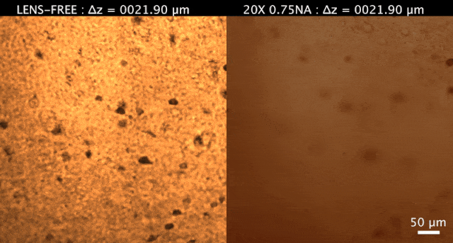
 Lens-free holographic microscopy (left) and scanning optical microscope (right) of a 200-micron thick mouse brain sampleZHANG ET AL., SCI ADV, 3:E1700553, 2017. A novel imaging technique allows researchers to peer through thick, three-dimensional tissues using a holographic microscope and economical, easily transported tools, according to a study published today (August 11) in Science Advances. The tissue preparation and imaging protocol is meant to serve as an alternative to costlier techniques that aren’t easily accessible in resource-starved areas.
The researchers demonstrated the utility of their technique on a slice of a mouse brain, 200 microns (0.2 millimeters) in thickness. First, they made their tissue sample see-through using a tweaked version of the CLARITY tissue-clearing method, which removes lipids within tissue, then applied a stain to visualize brain cells. The researchers placed their sample near an image-sensing chip, which digitally acquired a focused, 3-D image. Traditional techniques, on the other hand, require lenses and cumbersome optical apparatuses.
They verified their reconstructed images by comparing them to those obtained by a scanning optical microscope. This test confirmed that the lens-free technique can be used to create a more-focused image.
“We believe that this CLARITY-enabled computational tissue imaging technique could find numerous applications in biomedical diagnosis ...


