Retinal degeneration is a leading cause of blindness globally and results from irreversible damage to photoreceptors and supporting cells including retinal pigment epithelium (RPE). Scientists have associated this deterioration with a variety of ocular diseases, such as retinitis pigmentosa (RP), which encompasses a group of inherited diseases, and age-related macular degeneration (AMD).

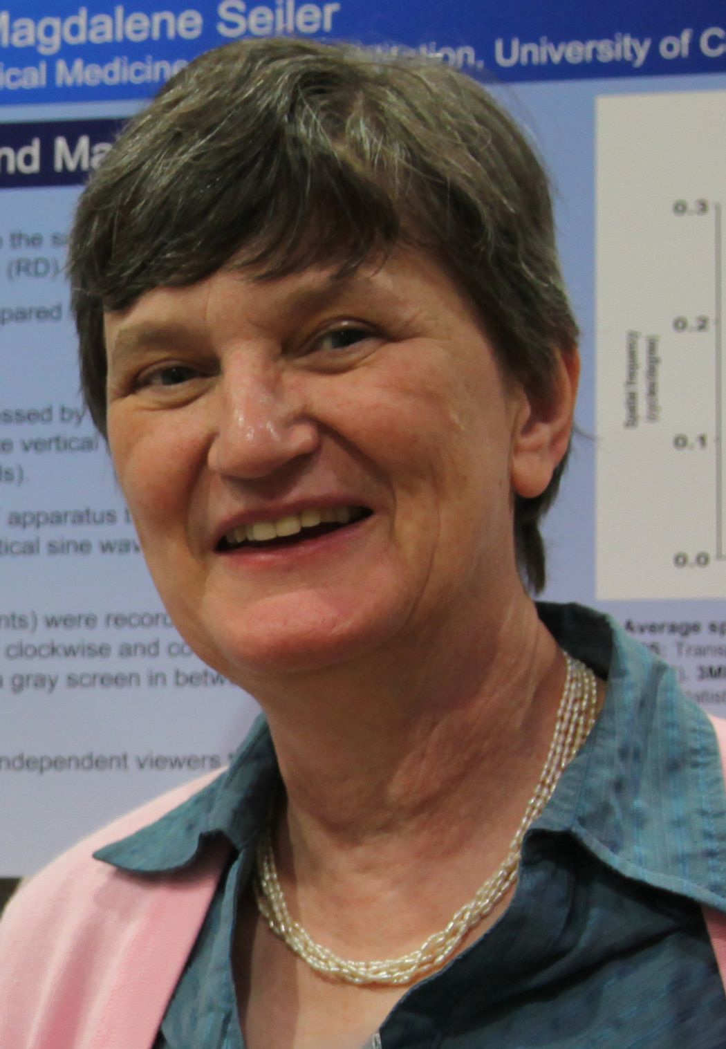
Magdalene Seiler and her team develop transplantable retinal organoid sheets to improve the vision of patients with retinal degeneration.
Magdalene Seiler
Magdalene Seiler, an ophthalmology researcher and neurobiologist at the University of California, Irvine (UCI), has spent over 30 years developing transplantation strategies to restore vision in patients affected by retinal degenerative diseases. She proposes that healthy photoreceptors can replace the degenerated or dead cells and form synaptic connections with the remaining host retina, thereby enhancing vision. In an interview with The Scientist, Seiler discussed the evolution of her retinal transplantation research and the obstacles her team must overcome to produce a clinical therapy.
What is the focus of your retinal transplantation research?
My team has developed a method to transplant retinal sheets into degenerating retinas.1 Because the retina has a very complex structure, it would be very difficult to get the photoreceptors to arrange in the correct orientation and organization if we dissociate the cells. By using sheet transplantation, we avoid this problem.
In our previous work, we isolated these retinal sheets from rat embryos and donated human fetal tissue, and demonstrated that transplantation of this tissue with its RPE improves the vision of several rat retinal degeneration models and patients with RP or AMD, respectively.2 However, there are ethical concerns and logistical problems with acquiring and using fetal tissue-based transplants.
So, we are now generating retinal organoid sheets, which provide us with essentially an unlimited source of tissue.3 We first differentiate human embryonic stem cells to form 3D spherical organoids. As these laminated structures contain photoreceptors on their surface, we then cut little pieces from their outermost layers and insert this tissue into the subretinal space of a rat. Like fetal retinal sheets, we have observed that these grafts also enhance the rat’s vision.
What challenges do scientists face when developing retinal organoid-based therapies?
Growing retinal organoids is a very labor- and material-intensive process, which ultimately can result in variations in the quality and quantity of the organoids obtained. This is a major problem for scientists employing these models in preclinical and clinical research. However, researchers can control the production and maintenance of retinal organoids by using uniform conditions. To accomplish this task, we developed a micro-millifluidic bioreactor to allow for continuous infusion of culture medium to the organoids.4 Normally, scientists must change half of the medium every two days. But using our method, we only have to change the medium reservoir once a week. This cuts down on labor and culture medium consumption. Additionally, this process is less stressful for the retinal organoids and it is much easier to maintain sterility, as there is less manipulation involved than standard culture.
Another major challenge to developing a clinical therapy using retinal organoids is maintaining their viability during long-distance shipping.5 We cannot produce the organoids in the same geographical location as the transplantation surgery and we needed to find a way to safely transport them under ideal conditions. Retinal organoids do not respond well to temperature fluctuations or vigorous shaking. So, we designed a shipping device that contains a portable incubator with a battery to maintain the temperature for one day.6 We also placed insulation material surrounding the incubator in the shipping crate to protect the organoids against bumps.
What are your next steps?
We would like to examine the connectivity between the retinal sheet transplant and the host cells. So, we are collaborating with the company Envigo to develop a transgenic retinal degenerated rat with fluorescently-labeled rod bipolar cells. In addition, we are working with David Lyon, also from UCI, to evaluate the success of the transplants by measuring vision recovery in the visual cortex, which is responsible for complex visual processing. Our ultimate long-term goal is to use retinal organoid sheets to develop a clinical therapy for patients with severe retinal degeneration. This will require a standardized protocol that consistently generates the same organoid every time, and further optimization of our bioreactor could help us meet this need.
This interview has been condensed and edited for clarity.
- Seiler MJ, Aramant RB. Intact sheets of fetal retina transplanted to restore damaged rat retinas. Invest Ophthalmol Vis Sci. 1998;39(11):2121-2131.
- Radtke ND, et al. Vision improvement in retinal degeneration patients by implantation of retina together with retinal pigment epithelium. Am J Ophthalmol. 2008;146(2):172-182.
- McLelland BT, et al. Transplanted hESC-derived retina organoid sheets differentiate, integrate, and improve visual function in retinal degenerate rats. Invest Ophthalmol Vis Sci. 2018;59(6):2586.
- Xue Y, et al. Retinal organoids on-a-chip: A 3D printed micro-millifluidic bioreactor for long-term retinal organoid maintenance. Lab Chip. 2021;21(17):3361.
- Lin B, et al. Survival and functional integration of human embryonic stem cell–derived retinal organoids after shipping and transplantation into retinal degeneration rats. Stem Cells Dev. 2024;33(9-10):201-213.
- Singh RK, et al. Development of a protocol for maintaining viability while shipping organoid-derived retinal tissue. J Tissue Eng Regen Med. 2020;14(2):388-394.


