 |
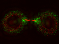
Image copyright Science/AAAS |
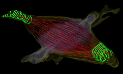 |
Image copyright Science/AAAS |
**__Related stories:__***linkurl:Top 7 papers in cell biology;http://www.the-scientist.com/news/display/57854/
[6th December 2010] *linkurl:New cell cycle complexities;http://www.the-scientist.com/blog/display/55659/
[23rd April 2009] *linkurl:Cell division rewinds;http://www.the-scientist.com/article/gateway/23551/
[1st June 2006] *linkurl:Related F1000 evaluations;http://f1000.com/search/evaluations?query=cell+division
[10th February 2011]


