Food's Afterlife

Meals left to mold develop colors, mycelia, and beads of digested juices, sparking the eye of an artist, and the slight concern of a mycologist.

Written byEdyta Zielinska
| 1 min read

Register for free to listen to this article
Listen with Speechify
0:00
1:00
Share

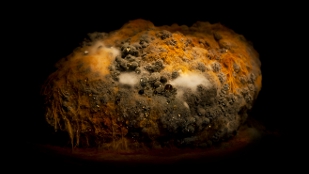
Mold growing on a rutabagaAFTERLIFE, HEIKKI LEIS

Those “science experiments” of thick fungus growing on coffee left for weeks on the desk, or sandwiches kept too long in the fridge are Estonian artist Heikki Leis's moldy muses. Afterlife is an exhibit of photographs taken of food Leis carefully let rot in his studio. It was on display last year at the AHHAA Science Centre in Estonia.

Wired Science blog invited mycologist Kathie Hodge of Cornell University to comment on the photos and try to spot the fungal species. “Most of the blue-grey molds are species of penicillium,” she told Wired, referring to photos that featured vegetables with splotches of the fuzzy blue species. “It seems that the artist may have a penicillium issue in his studio.” She picked out another familiar character ...

Interested in reading more?

Become a Member of

The Scientist Logo
Receive full access to more than 35 years of archives, as well as TS Digest, digital editions of The Scientist, feature stories, and much more!
Already a member? Login Here

Related Topics

Meet the Author

Share
Illustration of a developing fetus surrounded by a clear fluid with a subtle yellow tinge, representing amniotic fluid.
January 2026, Issue 1

What Is the Amniotic Fluid Composed of?

The liquid world of fetal development provides a rich source of nutrition and protection tailored to meet the needs of the growing fetus.

View this Issue
Skip the Wait for Protein Stability Data with Aunty

Skip the Wait for Protein Stability Data with Aunty

Unchained Labs
Graphic of three DNA helices in various colors

An Automated DNA-to-Data Framework for Production-Scale Sequencing

illumina
Exploring Cellular Organization with Spatial Proteomics

Exploring Cellular Organization with Spatial Proteomics

Abstract illustration of spheres with multiple layers, representing endoderm, ectoderm, and mesoderm derived organoids

Organoid Origins and How to Grow Them

Thermo Fisher Logo

Products

Brandtech Logo

BRANDTECH Scientific Introduces the Transferpette® pro Micropipette: A New Twist on Comfort and Control

Biotium Logo

Biotium Launches GlycoLiner™ Cell Surface Glycoprotein Labeling Kits for Rapid and Selective Cell Surface Imaging

Colorful abstract spiral dot pattern on a black background

Thermo Scientific X and S Series General Purpose Centrifuges

Thermo Fisher Logo
Abstract background with red and blue laser lights

VANTAstar Flexible microplate reader with simplified workflows

BMG LABTECH