

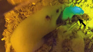
 One of the mouse pups born to a female who was implanted with a 3-D printed ovary, which contained follicles tagged with green fluorescent protein. NATURE COMMUNICATIONS, ISSN 2041-1723Adding to the already substantial list of organs that can be 3-D printed and function more or less normally, researchers have made mouse ovaries using the technique. Some of the live mouse they implanted with the prosthetic organs, after seeding them with egg-containing follicles, had normal young. The team of scientists, from Northwestern University, published the results in Nature Communications today (May 16).
The “landmark study” is a “significant advance in the application of bioengineering to reproductive tissues,” Mary Zelinski, a reproductive scientist at the Oregon National Primate Research Center in Beaverton who was not involved with the research, told Science.
The paper’s authors used a 3-D printer to lay down layers of gelatin, which is derived from collagen, on glass slides until they formed 15 mm x 15 mm scaffolds of varying density. They then inserted mouse follicles—balls of hormone-secreting cells encasing primordial ova—into the scaffolds and found after about a week that the scaffolds with smaller pores better supported follicles.
The researchers then inserted follicle-seeded, printed ovaries into seven female mice whose natural ovaries they had removed. The synthetic ovaries became vascularized within roughly a week, ...


