Pits Stopped

Editor’s choice in cell biology

Written byRuth Williams
| 2 min read

Register for free to listen to this article
Listen with Speechify
0:00
2:00
Share

Blocked Swapping
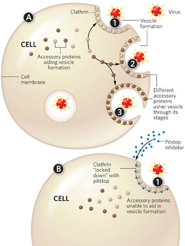
A. The clathrin lattice forms at the site of membrane invagination (1). Underneath the lattice accessory proteins come and go (2), each driving a different step in the invagination and vesicle-forming process (3). B. When a pitstop binds clathrin (1), it prevents these accessory proteins from binding to clathrin, and endocytosis stops.
PRECISION GRAPHICS

Clathrin-mediated endocytosis (CME)—by which cells ingest extracellular molecules and internalize cell surface proteins—is an essential process in practically all cells. Now, Volker Haucke of Freie Universität in Berlin and colleagues have identified two small molecules, which they named “pitstops” 1 and 2, that bind to clathrin’s terminal domain and stop CME. The inhibitors not only will help researchers elucidate clathrin’s molecular mechanisms, but could also serve as the basis for designing CME-preventing drugs for a variety of clinical applications.

“There are different ways to inhibit clathrin,” such as overexpressing or mutating accessory proteins, or using RNAi to deplete clathrin, says Ludger Johannes of the Institut Curie in Paris, who was not involved in the study. “But these all have limitations,” he adds. For example, with ...

Interested in reading more?

Become a Member of

The Scientist Logo
Receive full access to digital editions of The Scientist, as well as TS Digest, feature stories, more than 35 years of archives, and much more!
Already a member? Login Here

Related Topics

Meet the Author

  • ruth williams

    Ruth is a freelance journalist. Before freelancing, Ruth was a news editor for the Journal of Cell Biology in New York and an assistant editor for Nature Reviews Neuroscience in London. Prior to that, she was a bona fide pipette-wielding, test tube–shaking, lab coat–shirking research scientist. She has a PhD in genetics from King’s College London, and was a postdoc in stem cell biology at Imperial College London. Today she lives and writes in Connecticut.

    View Full Profile

Published In

Share
Illustration of a developing fetus surrounded by a clear fluid with a subtle yellow tinge, representing amniotic fluid.
January 2026

What Is the Amniotic Fluid Composed of?

The liquid world of fetal development provides a rich source of nutrition and protection tailored to meet the needs of the growing fetus.

View this Issue
Human-Relevant In Vitro Models Enable Predictive Drug Discovery

Advancing Drug Discovery with Complex Human In Vitro Models

Stemcell Technologies
Redefining Immunology Through Advanced Technologies

Redefining Immunology Through Advanced Technologies

Ensuring Regulatory Compliance in AAV Manufacturing with Analytical Ultracentrifugation

Ensuring Regulatory Compliance in AAV Manufacturing with Analytical Ultracentrifugation

Beckman Coulter Logo
Conceptual multicolored vector image of cancer research, depicting various biomedical approaches to cancer therapy

Maximizing Cancer Research Model Systems

bioxcell

Products

Refeyn logo

Refeyn named in the Sunday Times 100 Tech list of the UK’s fastest-growing technology companies

Parse Logo

Parse Biosciences and Graph Therapeutics Partner to Build Large Functional Immune Perturbation Atlas

Sino Biological Logo

Sino Biological's Launch of SwiftFluo® TR-FRET Kits Pioneers a New Era in High-Throughout Kinase Inhibitor Screening

SPT Labtech Logo

SPT Labtech enables automated Twist Bioscience NGS library preparation workflows on SPT's firefly platform