Scientists Create See-Through Hearts

A new tissue-clearing technique reveals blood vessel networks and collagen scaffolds of dog and human cardiac tissue.

Written byDiana Kwon
| 2 min read

Register for free to listen to this article
Listen with Speechify
0:00
2:00
Share

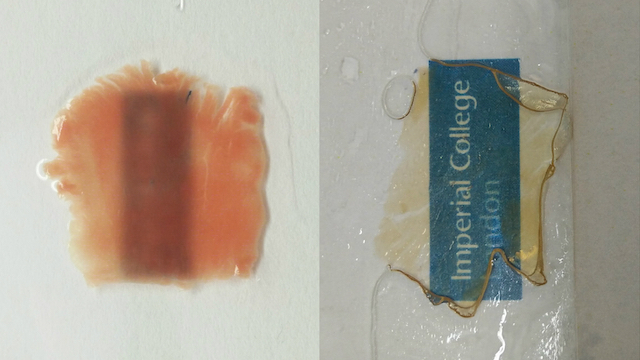
Tissue sample before (left) and after (right) treatmentFILLIPO PERBELLINIBy modifying CLARITY, an optical clearing technique first developed for brain tissue, to the heart, researchers at Imperial College London have created transparent cardiac tissue, according to a study published yesterday (July 12) in Scientific Reports.

CLARITY works by removing lipids from tissue, allowing light to penetrate. In recent years, researchers have applied this method various tissues, such as bone, and even to a whole mouse.

In the latest study, researchers combined CLARITY and DISCO (3-D imaging of solvent-cleared organs), another tissue-clearing technique, to create a new method, free-of-acrylamide SDS-based tissue clearing (FASTClear), to make both canine and human heart tissue transparent.

“Cardiac tissue is quite dense, using standard microscopy methods we are able to image the surface of the samples, about 20-30 microns from the surface,” coauthor Filippo Perbellin, a research associate at Imperial College London, says in a statement. “With more sophisticated methods we can achieve 50-80 microns depth but we were never able to image a whole myocardial ...

Interested in reading more?

Become a Member of

The Scientist Logo
Receive full access to more than 35 years of archives, as well as TS Digest, digital editions of The Scientist, feature stories, and much more!
Already a member? Login Here

Related Topics

Meet the Author

  • Diana is a freelance science journalist who covers the life sciences, health, and academic life. She’s a regular contributor to The Scientist and her work has appeared in several other publications, including Scientific American, Knowable, and Quanta. Diana was a former intern at The Scientist and she holds a master’s degree in neuroscience from McGill University. She’s currently based in Berlin, Germany.

    View Full Profile
Share
Illustration of a developing fetus surrounded by a clear fluid with a subtle yellow tinge, representing amniotic fluid.
January 2026, Issue 1

What Is the Amniotic Fluid Composed of?

The liquid world of fetal development provides a rich source of nutrition and protection tailored to meet the needs of the growing fetus.

View this Issue
Skip the Wait for Protein Stability Data with Aunty

Skip the Wait for Protein Stability Data with Aunty

Unchained Labs
Graphic of three DNA helices in various colors

An Automated DNA-to-Data Framework for Production-Scale Sequencing

illumina
Exploring Cellular Organization with Spatial Proteomics

Exploring Cellular Organization with Spatial Proteomics

Abstract illustration of spheres with multiple layers, representing endoderm, ectoderm, and mesoderm derived organoids

Organoid Origins and How to Grow Them

Thermo Fisher Logo

Products

Brandtech Logo

BRANDTECH Scientific Introduces the Transferpette® pro Micropipette: A New Twist on Comfort and Control

Biotium Logo

Biotium Launches GlycoLiner™ Cell Surface Glycoprotein Labeling Kits for Rapid and Selective Cell Surface Imaging

Colorful abstract spiral dot pattern on a black background

Thermo Scientific X and S Series General Purpose Centrifuges

Thermo Fisher Logo
Abstract background with red and blue laser lights

VANTAstar Flexible microplate reader with simplified workflows

BMG LABTECH