Updated Tissue-Clearing Protocol Extends Time Frame for Imaging

“Ultimate DISCO” uses a solvent that shrinks whole animals and preserves fluorescence for months.

Written byKerry Grens
| 1 min read

Register for free to listen to this article
Listen with Speechify
0:00
1:00
Share

YOUTUBE, ALI ERTURKA variety of techniques sweep away lipids and other opaque molecules to create see-through biological samples, including whole animals. Reporting in Nature Methods yesterday (August 22), scientists presented a tissue-clearing protocol that shrinks tissue to expand the limits of how big samples can be for imaging.

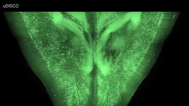
“We imaged the complete central nervous system of mice, and you can track individual cells several centimetres long that reach from the brain right through to the tip of the spinal cord,” study coauthor Ali Ertürk of the Ludwig Maximilian University of Munich in Germany told New Scientist.

Ertürk and colleagues built upon a procedure called 3-D imaging of solvent-cleared organs (DISCO). Although DISCO shrinks samples, it “quickly quenches endogenously expressed fluorescent proteins (with a half-life of a few days),” the authors point out in their report. They searched for solvents that could clear tissue without destroying fluorescence and settled upon diphenyl ether. Their recipe, called “ultimate DISCO,” maintains fluorescence for months.

“I love it,” Ingo Bechmann, a neuroscientist at the University of Leipzig, Germany, ...

Interested in reading more?

Become a Member of

The Scientist Logo
Receive full access to more than 35 years of archives, as well as TS Digest, digital editions of The Scientist, feature stories, and much more!
Already a member? Login Here

Related Topics

Meet the Author

  • kerry grens

    Kerry served as The Scientist’s news director until 2021. Before joining The Scientist in 2013, she was a stringer for Reuters Health, the senior health and science reporter at WHYY in Philadelphia, and the health and science reporter at New Hampshire Public Radio. Kerry got her start in journalism as a AAAS Mass Media fellow at KUNC in Colorado. She has a master’s in biological sciences from Stanford University and a biology degree from Loyola University Chicago.

    View Full Profile
Share
Illustration of a developing fetus surrounded by a clear fluid with a subtle yellow tinge, representing amniotic fluid.
January 2026, Issue 1

What Is the Amniotic Fluid Composed of?

The liquid world of fetal development provides a rich source of nutrition and protection tailored to meet the needs of the growing fetus.

View this Issue
Skip the Wait for Protein Stability Data with Aunty

Skip the Wait for Protein Stability Data with Aunty

Unchained Labs
Graphic of three DNA helices in various colors

An Automated DNA-to-Data Framework for Production-Scale Sequencing

illumina
Exploring Cellular Organization with Spatial Proteomics

Exploring Cellular Organization with Spatial Proteomics

Abstract illustration of spheres with multiple layers, representing endoderm, ectoderm, and mesoderm derived organoids

Organoid Origins and How to Grow Them

Thermo Fisher Logo

Products

Brandtech Logo

BRANDTECH Scientific Introduces the Transferpette® pro Micropipette: A New Twist on Comfort and Control

Biotium Logo

Biotium Launches GlycoLiner™ Cell Surface Glycoprotein Labeling Kits for Rapid and Selective Cell Surface Imaging

Colorful abstract spiral dot pattern on a black background

Thermo Scientific X and S Series General Purpose Centrifuges

Thermo Fisher Logo
Abstract background with red and blue laser lights

VANTAstar Flexible microplate reader with simplified workflows

BMG LABTECH