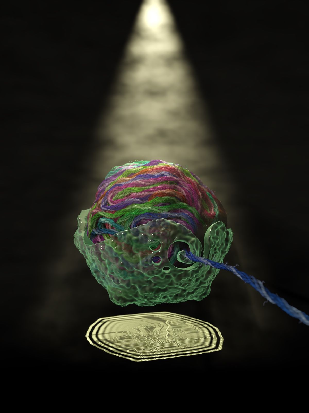
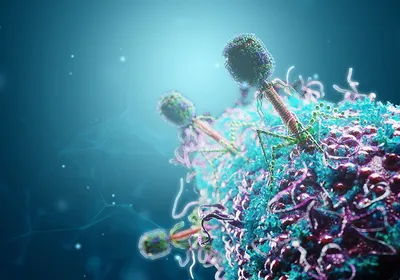
A virus may be microscopic, but it contains thousands of nucleic acid bases strategically packaged into a protein shell. Knowing how the virus organizes these vast information stores in a compact space is the key to understanding viral structure and designing better defenses against pathogenic viruses.
Peering into the viral protein shell, or capsid, is challenging. Typical structure discerning techniques such as cryo-electron microscopy can’t capture the varying configurations of genetic material in each virus. Back in 2010, Aleksei Aksimentiev, a biophysicist at the University of Illinois Urbana-Champaign, had an idea for computationally simulating a virus’s structure. However, computational methods were simply not sophisticated enough at the time.
“It was always at the back of our minds, and then, we made a breakthrough in terms of methodology,” Aksimentiev said.
Now, 14 years later, in a study published in Nature, his team reported using a new computational approach to simulate the individual atoms of a virus that is packed with nucleic acids.1 They used this method to study the HK97 bacteriophage and proposed the first structure for the virus.
A few years ago, Aksimentiev’s team developed a method for mapping out complex DNA configurations by computationally simulating them at multiple resolutions.2 They start at a coarse resolution, like a fuzzy image, and then on each iteration, they increase the level of detail in the simulated DNA structure.

In their new study, the researchers used this approach to computationally model the virus and its DNA during viral assembly. With prior experimental data such as the structure of the capsid and the force of the motor that loads DNA into the virus as a starting point, they simulated the behavior of each of the 26 million atoms during the chaotic process of loading DNA into the capsid. This was no small task; each simulation took anywhere between three months and one year to run, even on very powerful computers.
According to Eric May, a structural biologist at the University of Connecticut who was not involved in this study, the simulations provided unprecedented insights into the dynamics between the genome, the capsid, and other molecules in the virus that may be missed by experimental methods that can only obtain the average structure across many particles. “This computational approach doesn't have that kind of limitation,” he said. “We know the protein components already, but now seeing the genomic information in full atomic detail is very exciting.”
For example, the researchers predicted that DNA is packaged into the capsid through a method called loop extrusion, where proteins force the DNA into hairpin configurations.3 Aksimentiev was surprised to see the diversity of genomic configurations produced by the simulations.
“We intuitively would think each configuration could be different, but what was surprising to us was the scale at which the structures were different,” Aksimentiev said. “If you look at the individual viral particles, they are different by the global configuration, which was introduced by the varied packaging process.”
Matthias Wolf, a structural biologist at the Okinawa Institute of Science and Technology who was not involved in this study, said that this addresses a long standing question about how viruses organize their genomes. However, he noted that the study lacked experimental validation of the predicted structures.
Aksimentiev thinks that they can improve the simulation to account for more physical forces and be less reliant on experimental input data. His group is also running simulations of other viruses that are more complex. May thinks that it will be critical to apply this model to pathogenic viruses such as HIV and SARS-CoV-2, even though they are harder to model because of their RNA genomes. “It would be interesting to see [the researchers] try to move in the direction of systems of great public health importance,” he said. “Also understanding the stages of viral infection: how is the structure of a virus different when it enters the cell? How is the genome getting released from a virus?”
Aksimentiev is optimistic that the simulations will extend to more complex viruses, including RNA viruses, by incorporating more targeted experimental data. His eye is also on an even loftier goal: simulating an entire cell. “It's probably not coming soon, but that's kind of the Holy Grail,” he said.
References
1. Coshic K, et al. The structure and physical properties of a packaged bacteriophage particle. Nature. 2024;627(8005):905-914.
2. Maffeo C, Aksimentiev A. MrDNA: A multi-resolution model for predicting the structure and dynamics of DNA systems. Nucleic Acids Res. 2020;48(9):5135–5146.
3. Ganji M, et al. Real-time imaging of DNA loop extrusion by condensin. Science. 2018;360(6384):102-105.


