

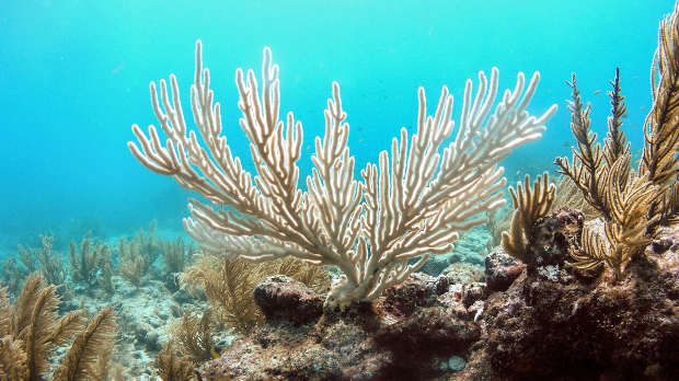
 Bleached colony of the soft coral bent sea rod WIKIMEDIA, KELSEY ROBERTS, USGSWith El Niño subsiding, Australian authorities are assessing the damage from this year’s bleaching event, which impacted an estimated 93 percent of the 3,000 or so reefs that comprise the Great Barrier Reef (GBR). “We’re in the tail-end period, where we’re waiting for the final mortality total to unfold,” Terry Hughes of the ARC Centre of Excellence for Coral Reef Studies at James Cook University told The Scientist at the International Coral Reef Symposium, which is ongoing this week in Honolulu, Hawaii.
And so far, the results are not looking good. “The corals we’re looking at are almost falling apart. They’re the sickest we’ve ever seen,” said Bill Leggat, associate professor with the ARC Centre of Excellence for Coral Reef Studies. “If we couple that with what we’re seeing with the visual surveys, with the number of corals impacted and the severity of the stress and the damage that we’re seeing, then it gets to be quite a scary story.”
When corals are stressed, they lose the symbiotic algae that live in their tissues, leading to a loss of color called bleaching. But not all the algae have to leave for the color to be lost; corals can bleach to different degrees, even if they look completely ...


.jpg)