As the foundation for connective tissues like tendons, bones, cartilage, and skin, collagen is the most abundant protein in the human body. It serves as the body’s scaffolding, supporting cells as they grow and function. Despite its importance for life, collagen has an unusual feature: It is not stable at body temperature.
Fascinated by this contradiction, Nancy Forde, a biophysicist at Simon Fraser University, and her colleagues investigated the features in collagen type IV that play a role in its stability. Their findings, published in the Proceedings of the National Academy of Sciences, identified cysteines as crucial chemical staples that help collagen IV remain intact.1 A better understanding of these structural features can provide insights into chemically altered collagen associated with connective tissue disorders.
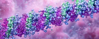
When stable, collagen IV has a triple-helix structure consisting of three strands twisted together like a thread. At higher temperatures, such as body temperature, these threads unravel into random coils. Forde likened collagen IV’s appearance to “a little lollipop,” with one globular region called the non-collagenous 1 (NC1) domain at the C-terminus and a thread-like collagenous domain extending from this to the N-terminus.
On a molecular level, this triple-helical structure is made up of repeating glycine-X-Y sequences, with proline and hydroxyproline typically filling the X and Y spots. But collagen IV has interruptions in this pattern where glycine is missing from every third amino acid. These breaks boost flexibility but also slow down its folding into the triple helix.

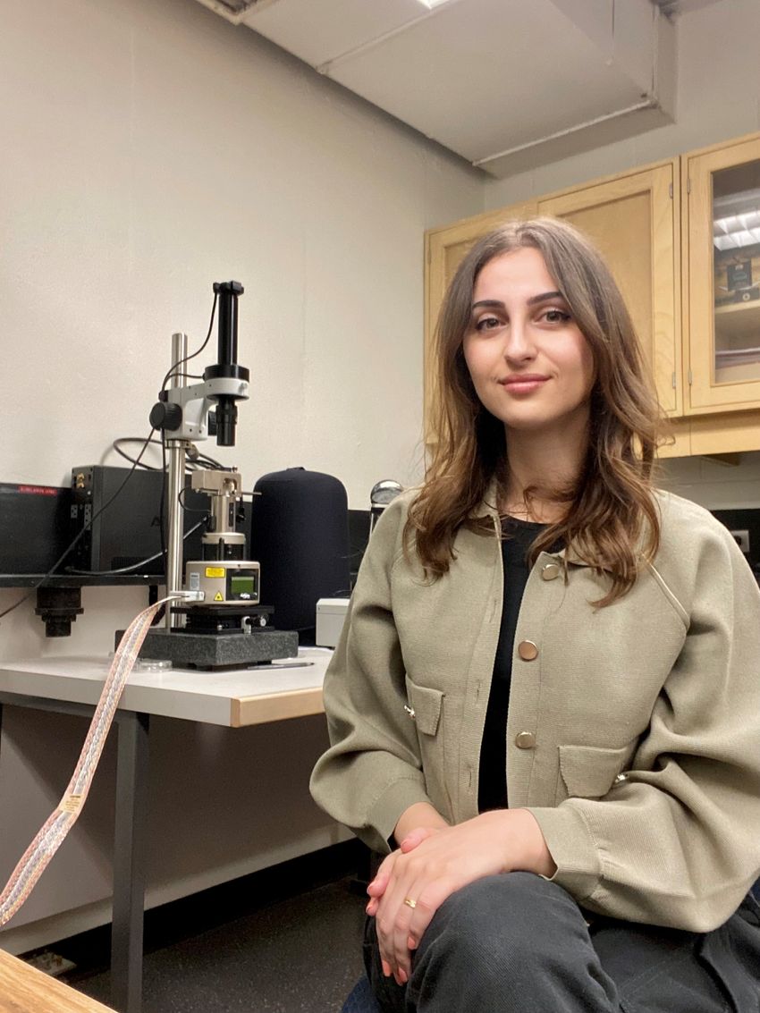
As a graduate student in Forde’s group, Alaa Al-Shaer investigated the perplexing mystery behind collagen IV’s thermal instability.
Simon Fraser University
To study individual collagen IV strands, Forde’s then graduate student Alaa Al-Shaer used atomic force microscopy to map the conformations of the protein. The researchers hypothesized that these interruptions might form an area of micro-unfolding that would be one of the first areas to unravel under heat and appear as extended, more flexible bulges. Al-Shaer assessed collagen IV’s conformation under dry conditions at 4°C, 22°C (room temperature), and 37°C (body temperature). The threads unraveled significantly at body temperature but apparently not within the collagenous domain in the spots the team expected. Instead, Forde remarked, “There was only one location in the whole sequence where we noticed a significant mechanical change [prior to unfolding].” This region of interest was just 200 to 250nm from the NC1 domain, approximately two-thirds of the way from the NC1 domain to the N-terminus.
“Atomic force microscopy has never been applied to collagen in this fashion [of folding and unfolding],” said Abhishek Jalan, a biochemist at the University of Bayreuth, who was not involved in the study. “It’s a very remarkable study in that respect.”
While investigating the area N-terminal to the changed region, the researchers noted that it seemed to mark the position of last resistance. Collagen IV retained its structure N-terminal to this region, but C-terminal to this point, the chains unraveled.
They observed that each of the three collagen chains contained two cystine residues positioned just one to two amino acids apart. These closely spaced cystines paired to form disulfide bonds, acting like chemical staples, that helped hold the chains together. In total, six cystine residues formed three disulfide bonds, creating what the researchers called a “cystine knot.”
This knot plays a crucial role in stabilizing collagen structure. At body temperature, the collagen IV was prone to unraveling up until the knot, but when it cooled, the collagen started refolding, pulling the coils back together. In contrast, collagen IV strands lacking these cystine staples fell apart much more quickly and couldn't reassemble. During these refolding events, the researchers were also surprised to see that as collagen IV reassembled overnight, it refolded from the N- to C-terminus direction. These findings contradict the current paradigm, which holds that collagen folds from the C- to the N-terminus.2
Jalan found the observed refolding direction perplexing. “The NC1 domains have a strong tendency to self-associate,” he explained. Because of this, he questioned how cystine residues, which would require close proximity to form stabilizing disulfide bonds and the subsequent knot formation, could drive the refolding process instead of the NC1 domain. However, Jalan remarked that further experimentation is needed to explore this proposed paradigm shift.
Although collagen is found across the animal kingdom, different types of collagens evolved at different times. Jalan noted, “One argument is that there is not [a single] folding mechanism that binds all different types of collagens. It’s a folding puzzle and unless all the pieces are together, a complete picture cannot emerge.” The cystine knot is one piece, while other elements—such as protein chaperones and interchain salt bridges—may also play critical roles in the protein’s stability.
Building on this work, Forde’s group plans to apply this approach to other collagens, such as procollagen I, which also features a globular end, and to explore how mutations and other chemical changes in collagens can lead to disease.
- Al-Shaer A, Forde NR. Decoding collagen's thermally induced unfolding and refolding pathways. Proc Natl Acad Sci USA. 2025;122(20):e2420308122.
- Engel J, Prockop DJ. The zipper-like folding of collagen triple helices and the effects of mutations that disrupt the zipper. Annu Rev Biophys Biophys Chem. 1991;20:137-152.


