Dozens of Researchers Exposed to Anthrax

As many as 75 scientists at the US Centers for Disease Control and Prevention may have come in contact with live Bacillus anthracis, the bacteria that cause anthrax.

Written byBob Grant
| 2 min read

Register for free to listen to this article
Listen with Speechify
0:00
2:00
Share

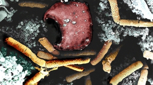
Color-enhanced scanning electron micrograph shows rod-shaped Bacillus anthracis (yellow) and a red blood cell (red) in a monkey’s spleen.NIAID, ARTHUR FRIEDLANDERApproximately 75 researchers working at the US Centers for Disease Control and Prevention (CDC) in Atlanta may have been exposed to live anthrax bacteria when the microbes were sent to laboratories that were not equipped to safely handle the pathogen, the federal agency announced Thursday (June 19).

CDC scientists were studying the bacteria in a high-level biosecurity lab and then transferred the samples to three lower-security labs without first properly inactivating the live microbes. Workers in those labs handled the samples without using personal protective equipment, such as gloves. The procedural lapse happened between June 6 and June 13, and CDC spokesperson Thomas Skinner told The New York Times that while none of the researchers exposed to the bacteria are showing symptoms of infection, many of them are receiving prophylactic antibiotic treatment “out of an abundance of caution.”

CNN reported on Thursday afternoon that 54 CDC employees determined to have been in the labs or hallways at the time live anthrax was present have been seen by the agency’s Occupational Health Clinic. ...

Interested in reading more?

Become a Member of

The Scientist Logo
Receive full access to more than 35 years of archives, as well as TS Digest, digital editions of The Scientist, feature stories, and much more!
Already a member? Login Here

Related Topics

Meet the Author

  • From 2017 to 2022, Bob Grant was Editor in Chief of The Scientist, where he started in 2007 as a Staff Writer. Before joining the team, he worked as a reporter at Audubon and earned a master’s degree in science journalism from New York University. In his previous life, he pursued a career in science, getting a bachelor’s degree in wildlife biology from Montana State University and a master’s degree in marine biology from the College of Charleston in South Carolina. Bob edited Reading Frames and other sections of the magazine.

    View Full Profile
Share
Illustration of a developing fetus surrounded by a clear fluid with a subtle yellow tinge, representing amniotic fluid.
January 2026, Issue 1

What Is the Amniotic Fluid Composed of?

The liquid world of fetal development provides a rich source of nutrition and protection tailored to meet the needs of the growing fetus.

View this Issue
Skip the Wait for Protein Stability Data with Aunty

Skip the Wait for Protein Stability Data with Aunty

Unchained Labs
Graphic of three DNA helices in various colors

An Automated DNA-to-Data Framework for Production-Scale Sequencing

illumina
Exploring Cellular Organization with Spatial Proteomics

Exploring Cellular Organization with Spatial Proteomics

Abstract illustration of spheres with multiple layers, representing endoderm, ectoderm, and mesoderm derived organoids

Organoid Origins and How to Grow Them

Thermo Fisher Logo

Products

Brandtech Logo

BRANDTECH Scientific Introduces the Transferpette® pro Micropipette: A New Twist on Comfort and Control

Biotium Logo

Biotium Launches GlycoLiner™ Cell Surface Glycoprotein Labeling Kits for Rapid and Selective Cell Surface Imaging

Colorful abstract spiral dot pattern on a black background

Thermo Scientific X and S Series General Purpose Centrifuges

Thermo Fisher Logo
Abstract background with red and blue laser lights

VANTAstar Flexible microplate reader with simplified workflows

BMG LABTECH