Einstein’s Unusual Brain

Previously unreleased photographs show that Einstein’s brain had several unusual features that could explain his extraordinary cognitive abilities.

Written byDan Cossins
| 1 min read

Register for free to listen to this article
Listen with Speechify
0:00
1:00
Share

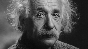
Wikimedia, Library of CongressPhotographs of Albert Einstein’s brain, taken shortly after his death but never previously released to the public, have revealed several unusual topographic features that may explain why the physicist was so clever, according to a study out last week (November 16) in Brain.

After Einstein died in 1955, pathologist Thomas Harvey removed and preserved the physicist’s brain. He took dozens of black and white photos from various angles before the brain was cut into 240 blocks, from which histological slides were prepared and sent to leading neuropathologists. Most of this material seems to have been lost over the years, but in 2010 what remains of Harvey’s private collection was given to the US Army's National Museum of Health and Medicine in Silver Spring, Maryland.

Having obtained 14 photographs from the collection, anthropologist Dean Falk from Florida State University and colleagues compared the folds and grooves of Einstein’s brain to those of 85 brains photographed for other studies. Although Einstein’s brain was average in size, they noticed patterns rarely ...

Interested in reading more?

Become a Member of

The Scientist Logo
Receive full access to more than 35 years of archives, as well as TS Digest, digital editions of The Scientist, feature stories, and much more!
Already a member? Login Here

Related Topics

Meet the Author

Share
Illustration of a developing fetus surrounded by a clear fluid with a subtle yellow tinge, representing amniotic fluid.
January 2026, Issue 1

What Is the Amniotic Fluid Composed of?

The liquid world of fetal development provides a rich source of nutrition and protection tailored to meet the needs of the growing fetus.

View this Issue
Skip the Wait for Protein Stability Data with Aunty

Skip the Wait for Protein Stability Data with Aunty

Unchained Labs
Graphic of three DNA helices in various colors

An Automated DNA-to-Data Framework for Production-Scale Sequencing

illumina
Exploring Cellular Organization with Spatial Proteomics

Exploring Cellular Organization with Spatial Proteomics

Abstract illustration of spheres with multiple layers, representing endoderm, ectoderm, and mesoderm derived organoids

Organoid Origins and How to Grow Them

Thermo Fisher Logo

Products

Brandtech Logo

BRANDTECH Scientific Introduces the Transferpette® pro Micropipette: A New Twist on Comfort and Control

Biotium Logo

Biotium Launches GlycoLiner™ Cell Surface Glycoprotein Labeling Kits for Rapid and Selective Cell Surface Imaging

Colorful abstract spiral dot pattern on a black background

Thermo Scientific X and S Series General Purpose Centrifuges

Thermo Fisher Logo
Abstract background with red and blue laser lights

VANTAstar Flexible microplate reader with simplified workflows

BMG LABTECH