
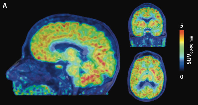
 [11C]Martinostat images averaged from 60 to 90 minutes after radiotracer injection from a representative subject overlaid on anatomical magnetic resonance imagesH.-Y. WEY ET AL., SCIENCE TRANSLATIONAL MEDICINEScientists at Massachusetts General Hospital and Harvard Medical School have—for the first time—imaged epigenetic regulation of gene expression in the living human brain. The team used positron emission tomography (PET) with an imaging probe, called [11C]Martinostat, that bound to particular histone deacetylases (HDACs), finding increased HDAC levels in cortical gray matter compared with white matter. The results were published yesterday (August 10) in Science Translational Medicine.
Gene transcription–regulating HDACs play a role in neurological disorders such as Alzheimer’s disease. The team applied its modified PET imaging approach on eight healthy volunteers, monitoring where the HDACs were switching off brain gene expression over time.
“It’s a good start for determining which epigenetic processes occur in the brain,” Isabelle Mansuy of the University of Zurich, Switzerland, who was not involved in the work told New Scientist.
“I’m hoping these colorful maps let us compare healthy brains with the brains of people with schizophrenia, Alzheimer’s, and other diseases,” Massachusetts General’s Jacob Hooker, who led the study, told STAT News.
“Just because we can see where HDACs are working doesn’t automatically mean we understand how to interpret the signals,” he told The Verge. “The biggest ...


