How Epstein-Barr Virus Hijacks B Cells To Cause Blood Cancer

Scientists describe a mechanism by which EBV controls the expression of two genes, leading B cells to replicate out of control and avoid apoptosis.

Written byJoshua A. Krisch
| 3 min read

Register for free to listen to this article
Listen with Speechify
0:00
3:00
Share

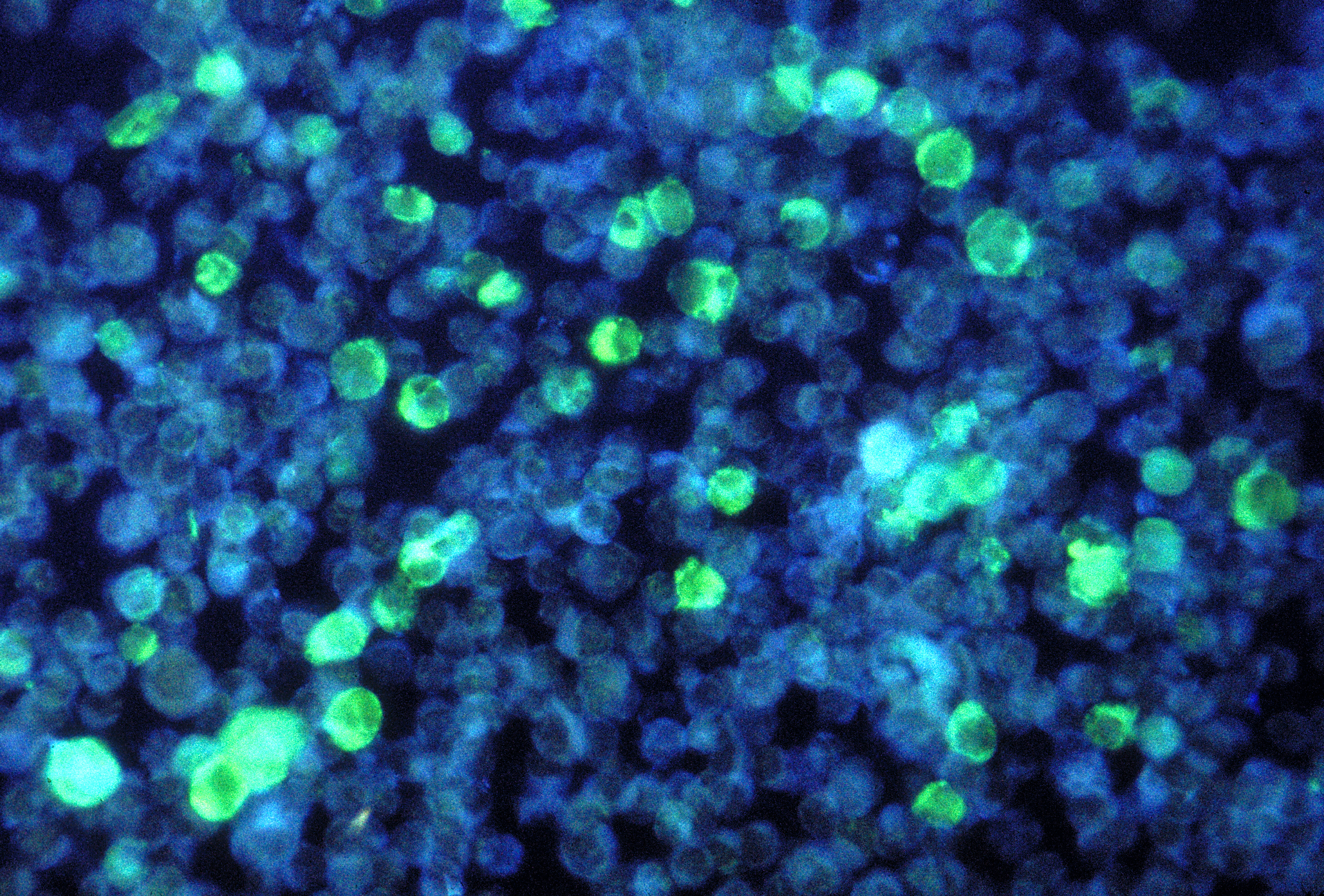
Leukemia cells containing Epstein-Barr VirusCDC, PAUL M. FEORINO

When Epstein-Barr virus (EBV) causes cancer, it usually does so by hijacking two cellular mechanisms. First, the virus deregulates MYC so that B cells can rapidly replicate. Then, the virus overrides the B cell’s built-in failsafe—programed cell death—by silencing an apoptotic gene, BCL2L11 (Bim). Now, in a November 10 study published in eLIFE, researchers describe one mechanism by which EBV controls the expression of these genes.

The two genes are “really important in cancer development,” said coauthor Michelle West, a biochemist at the University of Sussex, U.K. “We know the virus produces proteins that control these genes. We were interested in understanding how it turns on MYC and switches off another gene called BCL2L11.”

EBV, one of the first oncoviruses discovered, infects more than 90 ...

Interested in reading more?

Become a Member of

The Scientist Logo
Receive full access to more than 35 years of archives, as well as TS Digest, digital editions of The Scientist, feature stories, and much more!
Already a member? Login Here

Meet the Author

Share
Illustration of a developing fetus surrounded by a clear fluid with a subtle yellow tinge, representing amniotic fluid.
January 2026, Issue 1

What Is the Amniotic Fluid Composed of?

The liquid world of fetal development provides a rich source of nutrition and protection tailored to meet the needs of the growing fetus.

View this Issue
Skip the Wait for Protein Stability Data with Aunty

Skip the Wait for Protein Stability Data with Aunty

Unchained Labs
Graphic of three DNA helices in various colors

An Automated DNA-to-Data Framework for Production-Scale Sequencing

illumina
Exploring Cellular Organization with Spatial Proteomics

Exploring Cellular Organization with Spatial Proteomics

Abstract illustration of spheres with multiple layers, representing endoderm, ectoderm, and mesoderm derived organoids

Organoid Origins and How to Grow Them

Thermo Fisher Logo

Products

Brandtech Logo

BRANDTECH Scientific Introduces the Transferpette® pro Micropipette: A New Twist on Comfort and Control

Biotium Logo

Biotium Launches GlycoLiner™ Cell Surface Glycoprotein Labeling Kits for Rapid and Selective Cell Surface Imaging

Colorful abstract spiral dot pattern on a black background

Thermo Scientific X and S Series General Purpose Centrifuges

Thermo Fisher Logo
Abstract background with red and blue laser lights

VANTAstar Flexible microplate reader with simplified workflows

BMG LABTECH