Infographic: Dialing Up Dopamine

Synaptic vesicles can dynamically adjust neurotransmitter content in response to neuronal firing.

Written byCatherine Offord
| 2 min read

Register for free to listen to this article
Listen with Speechify
0:00
2:00
Share
© EVAN OTO/SCIENCE SOURCE
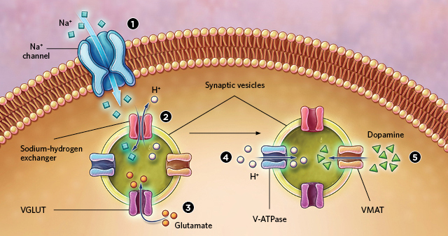
Researchers have found that synaptic vesicles releasing dopamine across neuronal synapses in fruit flies and mice can dynamically adjust the neurotransmitter content in response to neuronal firing. And they propose a mechanism to explain how. When the axon terminal depolarizes, sodium ions flow into the cell (1). The increased sodium ion concentration activates sodium-hydrogen exchangers in the vesicle membrane that transport one sodium ion into the vesicle for every proton out (2). This action increases the difference in electrical charge across the vesicle membrane, activating a transporter protein, VGLUT, which pumps another neurotransmitter, negatively charged glutamate, into the vesicle (3). The resulting buildup of internal negative charge triggers the pumping of more protons into the vesicle, increasing the pH gradient across the membrane (4). Finally, this drop in pH inside the vesicle triggers the loading of more dopamine into the vesicles via the VMAT protein (5).
Interested in reading more?

Become a Member of

The Scientist Logo
Receive full access to more than 35 years of archives, as well as TS Digest, digital editions of The Scientist, feature stories, and much more!
Already a member? Login Here

Meet the Author

  • After undergraduate research with spiders at the University of Oxford and graduate research with ants at Princeton University, Catherine left arthropods and academia to become a science journalist. She has worked in various guises at The Scientist since 2016. As Senior Editor, she wrote articles for the online and print publications, and edited the magazine’s Notebook, Careers, and Bio Business sections. She reports on subjects ranging from cellular and molecular biology to research misconduct and science policy. Find more of her work at her website.

    View Full Profile
Share
Illustration of a developing fetus surrounded by a clear fluid with a subtle yellow tinge, representing amniotic fluid.
January 2026

What Is the Amniotic Fluid Composed of?

The liquid world of fetal development provides a rich source of nutrition and protection tailored to meet the needs of the growing fetus.

View this Issue
Human-Relevant In Vitro Models Enable Predictive Drug Discovery

Advancing Drug Discovery with Complex Human In Vitro Models

Stemcell Technologies
Redefining Immunology Through Advanced Technologies

Redefining Immunology Through Advanced Technologies

Ensuring Regulatory Compliance in AAV Manufacturing with Analytical Ultracentrifugation

Ensuring Regulatory Compliance in AAV Manufacturing with Analytical Ultracentrifugation

Beckman Coulter Logo
Skip the Wait for Protein Stability Data with Aunty

Skip the Wait for Protein Stability Data with Aunty

Unchained Labs

Products

Sino Biological Logo

Sino Biological's Launch of SwiftFluo® TR-FRET Kits Pioneers a New Era in High-Throughout Kinase Inhibitor Screening

SPT Labtech Logo

SPT Labtech enables automated Twist Bioscience NGS library preparation workflows on SPT's firefly platform

nuclera logo

Nuclera eProtein Discovery System installed at leading Universities in Taiwan

Brandtech Logo

BRANDTECH Scientific Introduces the Transferpette® pro Micropipette: A New Twist on Comfort and Control