 CORPSE REMOVAL: An inner ear supporting cell (green) engulfs a dying hair cell (red) in the sensory epithelium of a mouse utricle.ELYSSA MONZACK The paper
E.L. Monzack et al., “Live imaging the phagocytic activity of inner ear supporting cells in response to hair cell death,” Cell Death Differ, doi:10.1038/cdd.2015.48, 2015.
Killer drugs
A number of commonly used medications can cause hearing loss by killing off cochlear hair cells, which translate sound waves into neural activity. To understand how they die, Lisa Cunningham and Elyssa Monzack of the National Institute on Deafness and Other Communication Disorders and colleagues turned to the utricle, a vestibular inner-ear structure involved with balance whose hair cells are very similar to those in the cochlea, which are notoriously resistant to culturing when mature.
Body bags
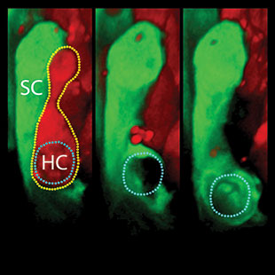
The team developed a method to watch hair cells of whole mouse utricles die in real time after exposure to the chemotherapy drug cisplatin or the antibiotic neomycin. In response to the latter, supporting cells, glia-like neighbors of hair cells, appeared to form a phagosome around the corpses and engulf them. “You can see two, three, sometimes four supporting cells advancing simultaneously on that hair cell corpse,” says Cunningham—which suggests that the dying cell is giving off a specific and local signal.
Spilled guts
In contrast, cisplatin-induced hair cell death provoked hardly any phagocytic reaction from supporting cells, about half of which themselves succumbed. Cunningham says this could have clinical implications if dead hair cells then spill their cytoplasmic contents into the tissue, which can result in an immune response that can cause even further damage.
Distress call
Mark Warchol of Washington University in St. Louis says it will be important to identify the signal supporting cells are responding to after neomycin treatment. “There’s some molecular signal by which the hair cell causes [supporting cells] to execute this process. And with cisplatin, they’re just not capable of doing it.”
Inner Ear Undertakers
Support cells in the inner ear respond differently to two drugs that kill hair cells.
Written byKerry Grens
Kerry served as The Scientist’s news director until 2021. Before joining The Scientist in 2013, she was a stringer for Reuters Health, the senior health and science reporter at WHYY in Philadelphia, and the health and science reporter at New Hampshire Public Radio. Kerry got her start in journalism as a AAAS Mass Media fellow at KUNC in Colorado. She has a master’s in biological sciences from Stanford University and a biology degree from Loyola University Chicago.
View Full Profile
Learn about our Editorial Policies.
| 2 min read

Register for free to listen to this article
Listen with Speechify
0:00
2:00
Share
Interested in reading more?

Become a Member of
Receive full access to digital editions of The Scientist, as well as TS Digest, feature stories, more than 35 years of archives, and much more!
Already a member? Login Here
Related Topics
Meet the Author

Kerry served as The Scientist’s news director until 2021. Before joining The Scientist in 2013, she was a stringer for Reuters Health, the senior health and science reporter at WHYY in Philadelphia, and the health and science reporter at New Hampshire Public Radio. Kerry got her start in journalism as a AAAS Mass Media fellow at KUNC in Colorado. She has a master’s in biological sciences from Stanford University and a biology degree from Loyola University Chicago.
View Full Profile
Published In
Share


