Making the Rounds

Circular RNA biogenesis occurs when RNA fragments are bent into closed loops of one or more exons and/or introns.

Written byCatherine Offord
| 2 min read

Register for free to listen to this article
Listen with Speechify
0:00
2:00
Share

Circular RNAs often form as the pre-mRNA molecule is processed into its final transcript via splicing, in which introns are removed and exons are linked together. Most circular RNAs are thought to be formed by a process called backsplicing, which joins one end of an exon to the other, or to an upstream exon, forming a circle. Researchers have recently published several models—not all of them necessarily mutually exclusive—to explain how different parts of the RNA molecule are brought into close proximity, encouraging backsplicing and turning a linear sequence into circular RNA.

THE SCIENTIST STAFF
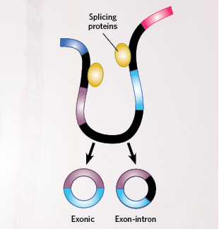
THE SCIENTIST STAFFIn a general backsplicing model, proteins assemble to form the spliceosome that processes transcribed RNA. But instead of splicing exons together in a linear sequence, they join the end of one exon to the beginning of the same exon or to an upstream exon. Below are three mechanisms that can drive this backsplicing

INTRON-PAIRING-DRIVEN CIRCULARIZATION

Complementary base pairs formed between long intronic sequences on different parts of the transcript bring together different splice sites on an RNA molecule, promoting backsplicing.

LARIAT-DRIVEN CIRCULARIZATION

Splicing proteins “skip” some ...

Interested in reading more?

Become a Member of

The Scientist Logo
Receive full access to more than 35 years of archives, as well as TS Digest, digital editions of The Scientist, feature stories, and much more!
Already a member? Login Here

Related Topics

Meet the Author

  • After undergraduate research with spiders at the University of Oxford and graduate research with ants at Princeton University, Catherine left arthropods and academia to become a science journalist. She has worked in various guises at The Scientist since 2016. As Senior Editor, she wrote articles for the online and print publications, and edited the magazine’s Notebook, Careers, and Bio Business sections. She reports on subjects ranging from cellular and molecular biology to research misconduct and science policy. Find more of her work at her website.

    View Full Profile
Share
Illustration of a developing fetus surrounded by a clear fluid with a subtle yellow tinge, representing amniotic fluid.
January 2026, Issue 1

What Is the Amniotic Fluid Composed of?

The liquid world of fetal development provides a rich source of nutrition and protection tailored to meet the needs of the growing fetus.

View this Issue
Skip the Wait for Protein Stability Data with Aunty

Skip the Wait for Protein Stability Data with Aunty

Unchained Labs
Graphic of three DNA helices in various colors

An Automated DNA-to-Data Framework for Production-Scale Sequencing

illumina
Exploring Cellular Organization with Spatial Proteomics

Exploring Cellular Organization with Spatial Proteomics

Abstract illustration of spheres with multiple layers, representing endoderm, ectoderm, and mesoderm derived organoids

Organoid Origins and How to Grow Them

Thermo Fisher Logo

Products

Brandtech Logo

BRANDTECH Scientific Introduces the Transferpette® pro Micropipette: A New Twist on Comfort and Control

Biotium Logo

Biotium Launches GlycoLiner™ Cell Surface Glycoprotein Labeling Kits for Rapid and Selective Cell Surface Imaging

Colorful abstract spiral dot pattern on a black background

Thermo Scientific X and S Series General Purpose Centrifuges

Thermo Fisher Logo
Abstract background with red and blue laser lights

VANTAstar Flexible microplate reader with simplified workflows

BMG LABTECH