

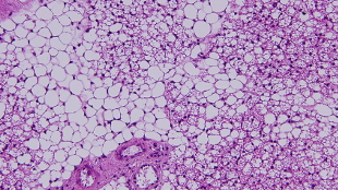
 Stained brown fat cells surrounded by white fat cellsSIEGFRIED USSARSince the discovery that not all fat cells are created equal, there has been a search for ways to reliably identify the so-called “good,” metabolically active, brown adipocytes, and the “bad,” relatively inert, white adipocytes. Thus far, biomarkers to distinguish brown from white fat have all been either intracellular or secreted proteins, which are not useful for whole-tissue or in vivo studies. Now, researchers have identified three cell-surface markers specific to adipocytes that are differentially expressed on white, brown, or third form of fat cell—beige—in both mice and in human tissue. The study is published today (July 30) in Science Translational Medicine.
“These markers look pretty selective and the key is that they are cell-surface markers, which will allow their use in a variety of applications in the future,” said Patrick Seale, an adipose tissue researcher at the University of Pennsylvania in Philadelphia who was not involved in the work. “I think these are going to be a really great resource for [researchers].”
White adipocytes store energy in the form of triglycerides, and depending on their location in the body, can have negative effects on health—contributing to insulin resistance, metabolic syndrome, and type 2 diabetes. Brown adipocytes are morphologically and functionally distinct, and are able to burn the energy stored as lipids while consuming glucose. “One of the major differences between brown and white fat is that ...


