Scattered throughout skeletal muscle, the muscle spindles are composed of connective tissue capsules containing bundles of specialized muscle fibers called intrafusal fibers. Wrapped around the middle of these fibers are the spiral terminals of a large sensory neuron, the type Ia axon. To one side of these spiral terminals, collectively called the primary endings, are the secondary endings, the terminals of a smaller sensory neuron, the type II axon. Both types of nerve endings serve as stretch sensors that send feedback about muscle contraction and length changes to the brain.
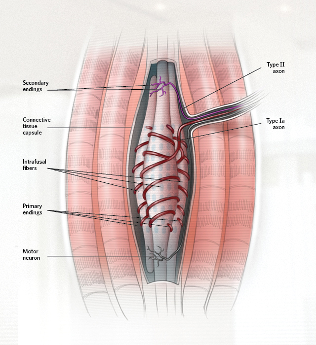 © CATHERINE DELPHIAAt rest, muscle spindles generate a trickle of nerve impulses. Stretching the muscle raises the number of nerve impulses from both the spindle’s primary and secondary endings. Impulses in the primary endings signal both the rate of change in muscle length and the length change itself. Primary endings are therefore both movement and position sensors. This sensitivity to the rate of the stretch makes the primary endings responsive to muscle vibration. The secondary endings respond only to the length change, making them position sensors; they are vibration-insensitive.
 © CATHERINE DELPHIA
Tendons attach muscles to bones. At the junction between muscle ...


