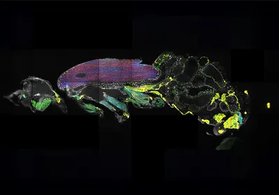
A color-annotated image of the muscles in the whole body of a fruit fly.
| 3 min read
Molecular cartography helped scientists map mRNA localization in the muscles and brains of adult Drosophila.

fluorescence microscopy

Lenses of a fluorescent microscope illuminate a sample on a microscope slide.

A Colorful Approach to Tracking Cellular Cargo

Microscopic image of a live amoeba.

Illuminating Specimens Through Live Cell Imaging

Charting a New Course Through the Injured Brain

An image of neurons, astrocytes, and other glial cells acquired using fluorescence microscopy.

Troubleshooting Fluorescence Microscopy Experiments

Dream Big and Achieve Real-Time Single Cell Imaging Without Camera Limitations

Sort What You See

Illustration of newly discovered mechanism allowing kinesin to “walk” down a microtubule. A green kinesin molecule with an attached yellow fluorophore is shown passing through a blue laser as it rotates step by step along a red and purple microtubule, fueled by blue ATP molecules that are hydrolyzed into orange ADP and phosphate groups.

High-Resolution Microscope Watches Proteins Strut Their Stuff

To flag neurons that have experienced genotoxic stress, researchers developed an in vivo sensor using an adeno-associated viral vector, called PRISM. Because a cell’s DNA damage response (DDR)—which activates in response to stressors such as environmental toxins or the buildup of misfolded proteins—also responds to invading pathogens, PRISM has an easier time transfecting cells whose damage response mechanisms are preoccupied with existing DNA damage. Once inside, the virus hijacks the neuron’s DNA replication machinery, which reverts an engineered frameshift mutation in the virus and thereby prompts the production of a fluorescent protein that can be observed via microscopy.

Infographic: DNA Damage Viewed with Unprecedented Clarity

Broken DNA

DNA Damage Viewed with Unprecedented Clarity

Microscopic image of nerves in the eye, a pathogen, and t cells

Science Snapshot: Eye Immunity

A scanning electron micrograph of a coculture of E. coli and Acinetobacter baylyi. Nanotubes can be seen extending from the E. coli.

What’s the Deal with Bacterial Nanotubes?

An illustration of a flask of bacteria, a weighted microscope slide, and two bacteria exchanging materials via nanotubes.

Infographic: Sources of Variation in Bacterial Nanotube Studies

Peering into the Cell

Infographic: Paraspeckle Form and Function

Organoids grown from a mouse’s colon

Caught on Camera

structures in a human cell

Deep Learning Algorithms Identify Structures in Living Cells

New Technique Captures Entire Fly Brain in 3D

Visualizing Gene Expression in Individual Cells in Thick Tissues

Image of the Day: Lego Microscopy

Image of the Day: Glowing Tick

Deep Learning Allows for Cell Analysis Without Labeling

Trending

Photo of John Calhoun crouches within his rodent utopia-turned-dystopia

Universe 25 Experiment

Raised hand holding a gold medal against blue sky.

What Is Gene Doping in Sports, and How Can Experts Detect It?

An MRI image of a human brain, with a glowing red section which indicates a tumor.

Why Are Breast Cancers That Metastasize to the Brain Hard to Treat?

Illustration of a pipette tip releasing a drop with a dollar sign into a test tube on a teal background.

Scientists Find Their Voices to Rally Support for Research

Multimedia

Close up concept art of PCR wells with colorful contents

Improving Cell Therapy Sterility Testing

sartorius logo
Conceptual photograph of connecting the dots in a pathway through route planning.

The Computer-Assisted Retrosynthesis Revolution

Merck
May digest 2025 cover
May 2025, Issue 1

Study Confirms Safety of Genetically Modified T Cells

A long-term study of nearly 800 patients demonstrated a strong safety profile for T cells engineered with viral vectors.

View this Issue
iStock

TaqMan Probe & Assays: Unveil What's Possible Together

Thermo Fisher Logo
Meet Aunty and Tackle Protein Stability Questions in Research and Development

Meet Aunty and Tackle Protein Stability Questions in Research and Development

Unchained Labs
Detecting Residual Cell Line-Derived DNA with Droplet Digital PCR

Detecting Residual Cell Line-Derived DNA with Droplet Digital PCR

Bio-Rad
How technology makes PCR instruments easier to use.

Making Real-Time PCR More Straightforward

Thermo Fisher Logo

Products

fujirebio-square-logo

Fujirebio Receives Marketing Clearance for Lumipulse® G pTau 217/ β-Amyloid 1-42 Plasma Ratio In-Vitro Diagnostic Test

The Scientist Placeholder Image

Biotium Launches New Phalloidin Conjugates with Extended F-actin Staining Stability for Greater Imaging Flexibility

Leica Microsystems Logo

Latest AI software simplifies image analysis and speeds up insights for scientists

BioSkryb Genomics Logo

BioSkryb Genomics and Tecan introduce a single-cell multiomics workflow for sequencing-ready libraries in under ten hours