The Evolution of Social Bees

Scientists describe the genetic changes associated with solitary-to-social transitions throughout bee evolution.

Written byRuth Williams
| 3 min read

Register for free to listen to this article
Listen with Speechify
0:00
3:00
Share

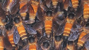
WIKIMEDIA, DR. AJAY BALACHANDRANSome bees are solitary, some live in small groups, and some live in colonies with many thousands of individuals. Studying the genomes of 10 bee species that represent these different living arrangements, scientists have now identified the genetic signatures of communal living. The results, published today (May 14) in Science, reveal that one key feature of increased sociality is an elaboration of gene regulation capacity.

“By doing this comparative analysis they show several clear changes associated with the evolution of the two eusocial groups of bees [including] changes in the regulation of gene expression,” said evolutionary ecologist Laurent Keller of the University of Lausanne in Switzerland who was not involved in the work. “Bees are highly social and we are highly social . . . so it’s interesting to see what are the mechanisms that lead to the evolution of such a complex system.”

While humans are highly social, cooperative creatures, people are not eusocial. A key feature of eusociality is the confinement of reproduction to select individuals—such as the queen in the case of bees. The switch from solitary living to eusociality, much like the transition from unicellular to ...

Interested in reading more?

Become a Member of

The Scientist Logo
Receive full access to more than 35 years of archives, as well as TS Digest, digital editions of The Scientist, feature stories, and much more!
Already a member? Login Here

Related Topics

Meet the Author

  • ruth williams

    Ruth is a freelance journalist. Before freelancing, Ruth was a news editor for the Journal of Cell Biology in New York and an assistant editor for Nature Reviews Neuroscience in London. Prior to that, she was a bona fide pipette-wielding, test tube–shaking, lab coat–shirking research scientist. She has a PhD in genetics from King’s College London, and was a postdoc in stem cell biology at Imperial College London. Today she lives and writes in Connecticut.

    View Full Profile
Share
Illustration of a developing fetus surrounded by a clear fluid with a subtle yellow tinge, representing amniotic fluid.
January 2026, Issue 1

What Is the Amniotic Fluid Composed of?

The liquid world of fetal development provides a rich source of nutrition and protection tailored to meet the needs of the growing fetus.

View this Issue
Skip the Wait for Protein Stability Data with Aunty

Skip the Wait for Protein Stability Data with Aunty

Unchained Labs
Graphic of three DNA helices in various colors

An Automated DNA-to-Data Framework for Production-Scale Sequencing

illumina
Exploring Cellular Organization with Spatial Proteomics

Exploring Cellular Organization with Spatial Proteomics

Abstract illustration of spheres with multiple layers, representing endoderm, ectoderm, and mesoderm derived organoids

Organoid Origins and How to Grow Them

Thermo Fisher Logo

Products

Brandtech Logo

BRANDTECH Scientific Introduces the Transferpette® pro Micropipette: A New Twist on Comfort and Control

Biotium Logo

Biotium Launches GlycoLiner™ Cell Surface Glycoprotein Labeling Kits for Rapid and Selective Cell Surface Imaging

Colorful abstract spiral dot pattern on a black background

Thermo Scientific X and S Series General Purpose Centrifuges

Thermo Fisher Logo
Abstract background with red and blue laser lights

VANTAstar Flexible microplate reader with simplified workflows

BMG LABTECH