Despite spanning about three billion base pairs, the human genome is wrapped up tight in a highly organized fashion in the nucleus. This coordinated structure, in part, enables the cell to regulate gene expression. One of the ways that cells accomplish this is through enhancer elements. These sequences are sometimes tens to hundreds of thousands of base pairs away from the genes they regulate, but by binding to transcription factors and physically looping the DNA, they bring these molecules into contact with a gene’s promoter.
In cancer, mutations and other genetic alterations can increase enhancer binding and subsequently drive gene expression.1 Although previous studies reported changes to 3D genetic architecture in tumors, researchers still don’t fully understand how these changes specifically affect enhancers and their role in gene regulation.2 While methods like RNA sequencing and assay for transposase-accessible chromatin using sequencing (ATAC-seq) inform researchers about DNA accessibility and use, these do not include structural information.3

As a graduate student at Stanford University, Kathryn Yost studied how gene regulation is altered in cancer, with a specific focus on the role of enhancer sequences.
Gretchen Ertl
“We know looking at just DNA sequencing, that there're a lot of structural rearrangements in the genomes of these cancers. But a lot of them are patient specific, so we don't actually know how those actually alter how enhancers are connected to target genes,” said Kathryn Yost, a cancer biologist and postdoctoral researcher at the Whitehead Institute. She added that since researchers usually map these data to the human reference genome, “We are a little bit blind.” She explained further that “in this particular tumor sample, maybe actually the genome is rearranged, and now this enhancer is actually not next to the gene that we think it is.”
As a graduate student in Howard Chang’s group at Stanford University, Yost and her colleagues addressed this shortcoming by leveraging The Cancer Genome Atlas (TCGA) and high-throughput conformational and sequencing techniques to map the 3D cancer genome.4 The findings, published in Nature Genetics, highlight how changes to enhancer binding influence gene expression in tumors.
Previously, methods like chromosome conformation capture with sequencing (Hi-C) and chromatin immunoprecipitation (ChIP)-based approaches that yielded architectural information either sacrificed specificity or required large numbers of cells.5,6 To overcome these limitations, Chang’s group developed HiChIP, which captures 3D structural information about the genome at specific loci and with smaller numbers of cells.7 It works by first crosslinking DNA-associated proteins, immunoprecipitating the crosslinked proteins, and then sequencing the bound DNA.
To find enhancer activity in cancer genomes, the team applied HiChip. Using histone H3 lysine 27 acetylation (H3K27ac) as a target for enhancer sequences, the researchers profiled 69 tumors from 15 different types of cancer available in TCGA to determine how enhancer binding activity changed across cancers. These samples included information from ATAC-seq, RNA-seq, and whole genome sequencing, allowing the team to combine their conformation data with that of accessible chromatin regions, gene expression, and mutations.
The researchers observed DNA loops with unique interactions of enhancer elements not previously reported. Some of these connections, for example those at the locus for the oncogene MYC, differed between cancer types; in one colon tumor, the researchers observed H34K27ac enrichment at the 5’ end of this gene, whereas in a liver tumor, they saw these marks in a 3’ regulatory region.
As they explored enhancer rewiring across 110 oncogenes, the team noticed three overall patterns: the same enhancer rewiring occurred around a gene across cancer types, a specific enhancer rewiring pattern only occurred in one cancer type, and—as in the example of MYC—enhancer rewiring patterns differed at the same gene in different cancers.
Next, the researchers investigated how enhancer rewiring and other DNA structural changes, such as gene duplications that result in higher gene expression, affected oncogene expression in their samples. Leveraging their HiChIP data with information from RNA-seq and whole genome sequencing, the researchers found that increased enhancer activity led to increased mRNA expression in more than 70 percent of oncogenes studied.
Jesse Dixon, a genome biologist at the Salk Institute of Biological Studies who was not involved in the study, said that this finding was informative. “One of the remarkable things about the way that they've done that is it actually shows that [gene duplications] may just be the tip of the iceberg,” he said. “Enhancer regulatory changes could actually be considerably more extensive in terms of the way that they’re driving [altered activation of] these types of genes.”
The team then explored altered enhancer activity in multiple different cell types in the tumor microenvironment of these different cancers by comparing HiChIP and ATAC-seq data. In one lung tumor sample, they observed increased interactions between enhancers and the promoter for the gene encoding programmed death-ligand 1 (PD-L1); data from ATAC-seq of the same sample showed that this region was accessible in myeloid cells as opposed to the tumor cells or other immune cells. Across cancer cell types, H3K27ac activity at this enhancer location correlated with PD-L1 expression and accessibility data pointed to it being specific to myeloid cells.

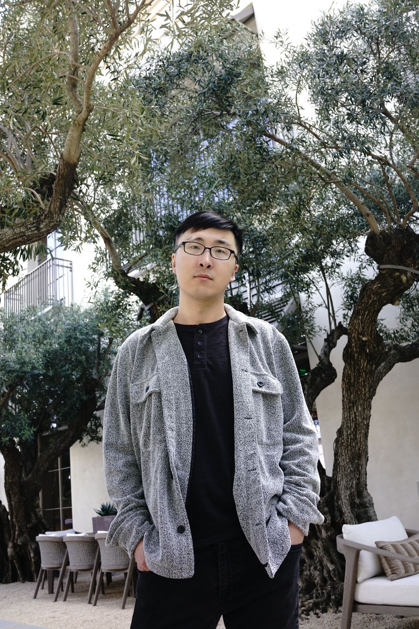
Yanding Zhao uses computational tools to study the cancer genome. He joined Chang’s group excited to study the role of enhancers in cancer.
Mengqing Tao
“This provides, I think, first, the mechanism of why tumor infiltrating myeloid [cells] have high PD-L1 expression,” said Yanding Zhao, a postdoctoral researcher and computational biologist in Chang’s group and study coauthor. Zhao added that these enhancers could be future cancer therapy targets.
Finally, the team investigated how mutations in noncoding regions influence gene expression and identified 7,517 expression-altering mutations in either promoter or enhancer regions. In stomach cancer samples, a mutation in a promoter for a gene involved in cell proliferation increased transcription factor binding to this region, increasing the production of mRNA at this site.
They also coupled their HiChIP data with whole genome sequencing information to demonstrate how certain structural rearrangements, such as translocations and extrachromosomal DNA, affected gene expression. The latter of these rearrangements created the highest number of new interactions between enhancers and promoters.
Dixon said that the study provides a valuable resource for studying cancer biology and identifying potential new drug targets based on these cancer-specific genome rearrangements. “What is nice about this is that I think that they've identified some kind of big picture regulatory phenomena that I think have not really been appreciated,” Dixon said. “This can be expanded to larger and larger cohorts.”
He added that although the study’s overall small sample size was a limitation, “I also think that that's a great opportunity.” He said, “There's a lot more that we can still learn in the future.”
- Flavahan WA, et al. Altered chromosomal topology drives oncogenic programs in SDH-deficient GISTs. Nature. 2019;575(7781):229-233.
- Johnstone SE, et al. Large-scale topological changes restrain malignant progression in colorectal cancer. Cell. 2020;182(6):1474-1489.e23.
- Buenrostro JD, et al. Transposition of native chromatin for fast and sensitive epigenomic profiling of open chromatin, DNA-binding proteins and nucleosome position. Nat Methods. 2013;10(12):1213-1218.
- Yost KE, et al. Three-dimensional genome landscape of primary human cancers. Nat Genet. 2025;57(5):1189-1200.
- Lieberman-Aiden E, et al. Comprehensive mapping of long-range interactions reveals folding principles of the human genome. Science. 2009;326(5950):289-293.
- Fullwood MJ, et al. An oestrogen-receptor-α-bound human chromatin interactome. Nature. 2009;462(7269):58-64.
- Mumbach MR, et al. HiChIP: Efficient and sensitive analysis of protein-directed genome architecture. Nat Methods. 2016;13(11):919-922.


