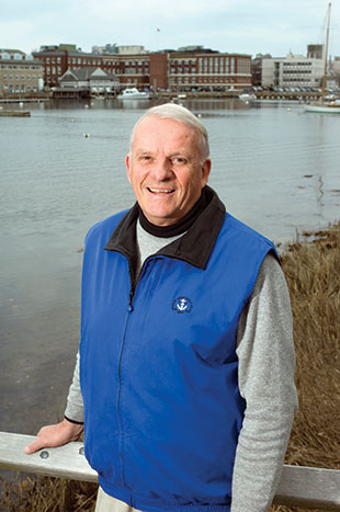
John E. Dowling
Gordon and Llura Gund Professor of Neurosciences Department of Molecular and Cellular Biology Harvard University
© TOM KLEINDINST/MBL
As a high school student in Providence, Rhode Island, John Dowling was not a good student. “I was doing too many other things, like playing sports, starting a school newspaper, and being a class officer,” he says. In tenth grade, he contracted polio and spent months recuperating. Not wanting him to lose the entire school year, his mother requested that Dowling’s teachers prepare lessons for him to do at home. “All of my teachers enthusiastically prepared the lessons except for my biology teacher, who wrote my mother that I was so hopeless in biology that I should drop the course.” Dowling gladly complied.

Dowling reconsidered his relationship with the subject during his undergraduate days at Harvard University, where he studied how vitamin A deficiency influences vision. He has...

Here, Dowling discusses how he helped revamp the biology curriculum at Harvard, pursued a PhD without knowing it, fished for laboratory supplies, and how, at age 79, he’s finally going to do a postdoctoral fellowship.

Dowling Debuts

Falling in love with biology. Dowling majored in biology at Harvard and planned to attend medical school. During his junior year, he took a biochemistry course taught by vision researcher and future Nobel laureate George Wald. “We first studied glycolysis, respiration, and photosynthesis, material that can be very dry, but George made it lively. He was a marvelous teacher. I can still recall his descriptions of Albert Szent-Györgyi’s famous experiments on muscle fibers: if you extract a frog or rabbit muscle with glycerol, you end up with a piece of inert tissue, but when you add back ATP, as long as the major proteins are intact, the muscles will contract. This excited me enormously, and still does. I thought it was really getting to the essence of life.”

First vision experiment. After learning about Wald’s discoveries of the role of vitamin A in vision, Dowling asked about working in his laboratory. “That’s where I fell in love with research,” says Dowling, His first research puzzle was to sort out why recovery from vitamin A deficiency is incomplete in humans who are placed on a vitamin A–containing diet. Prior studies in rats had suggested that prolonged vitamin A deficiency might cause degeneration of photoreceptors, so Dowling began with biochemical measurements on vitamin A–deficient rats. He found that levels of the visual pigment protein opsin decreased with time and the photoreceptor cells did indeed degenerate. Dowling learned how to record in vivo electroretinograms from the rats to understand what happens physiologically to their vision on a vitamin A–deficient diet. He observed that light sensitivity decreased logarithmically as the visual pigments in the retina declined. These experiments led to Dowling’s first publication—in 1958.

“I am finally closing the lab and embedding myself into the labs of Jeffrey Lichtman and Joshua Sanes to do a postdoctoral fellowship.”

Keeping the lab bench warm. In 1957, Dowling entered Harvard Medical School. “But Wald kept a lab bench for me, and on the odd afternoon during my first year at medical school I would come back and continue doing experiments. Then I spent the following summer working in the lab.” During that summer Dowling mapped the exchange of vitamin A and vitamin A aldehyde (retinal) between the retinal pigment epithelium and photoreceptor cells during light and dark adaptation as well as the relation between visual sensitivity and visual pigment levels during adaptation, showing that, just as with vitamin A deficiency, there is a logarithmic relationship between visual pigment levels and light sensitivity.

Permanent leave of absence. The following year, a member of the Wald lab learned of a study showing that when vitamin A–deficient rats consumed vitamin A acid (retinoic acid), the rats no longer showed signs of deficiency, even though no vitamin A could be detected in the animals’ tissues. Vitamin A in food is converted to retinol, its alcohol form, which is further metabolized to retinoic acid, but biological tissues have a hard time converting acids to aldehydes. “So we surmised that vitamin A acid could substitute for the somatic functions of vitamin A but not for the visual functions, which require retinal. I repeated the experiment, and the rats grew fine, but they became completely blind.”

Dowling decided to explore the functions of vitamin A and vitamin A acid in more detail—and to see if he could get the “research bug” out of his system. “I took a leave of absence from medical school in 1959, and I am still on that leave of absence 55 years later.” Dowling showed that while retinoic acid can indeed fulfill somatic tissue functions, it cannot be reduced to retinal, which is essential for vision. “With retinoic acid, we could study vitamin A deficiency confined to the eye—biochemically, electrophysiologically, and anatomically—and we showed that with long-term vitamin A deficiency, photoreceptors may be completely lost.”

Dowling Determined

PhD material. “Halfway through my leave of absence, Wald approached me about pursuing a PhD: ‘You’ve taken virtually all of the courses offered at Harvard in biology, have had two years of medical school, and you’ve done enough research to write a thesis. Why don’t you think about getting a PhD?’ Well, I never even thought about the possibility. This was long before there were any MD-PhD programs.” Dowling entered the PhD program in February 1960 and received his degree the following January.

Back to biology class. “I was bored silly when, as a freshman at Harvard, I had to endure introductory biology, which was taught as botany and then zoology, in two different semesters. Mostly it was just memorizing and had nothing about concepts,” says Dowling. So when Wald asked him to help teach a new introductory course that emphasized the commonalities shared by cells and organisms at the molecular level, Dowling jumped at the chance. And instead of going back to medical school as he had intended, Dowling accepted the offer of an assistant professor position at Harvard. “The biology department gave me a lab—I never did a postdoc.” Dowling helped teach the course for the three years he remained a junior faculty member at Harvard.

Making a move. In 1964, Dowling moved to the Wilmer Eye Institute at Johns Hopkins. While still at Harvard, Dowling had expanded from studying the low-light-sensitive rod photoreceptors that predominate in the rat retina to working on the ground squirrel retina, which contains mainly bright-light and color-sensitive cones. Dowling noticed that so-called horizontal cells in the retina received synaptic inputs from the photoreceptor cells. “The horizontal cells in those days were very much a mystery. This observation led to my main theme of research at Hopkins, working out the wiring of the retina and the physiology of the individual retinal neurons.”

Ahead of his time. Dowling quickly switched to the better-understood primate retina, working for five years with Brian Boycott to map the wiring of the retina’s various nerve cells, to identify its synapses, and to begin to understand the information flow within the eye. “Such mapping is very much in vogue now. The BRAIN Initiative announced by President Obama is exactly this idea—to anatomically reconstruct the nervous system. Well, we were trying to do this back in the 60s in the retina! The retina is a perfect neural structure to do reconstruction even though our methods, back then, were rudimentary compared to the large-scale computer reconstructions that can be done now.” Dowling and colleagues were the first to identify the synapses of the retinal neurons known as amacrine cells and to show how those synapses feed back onto retinal bipolar neuron terminals. Dowling also compared the primate retina to that of the frog, finding that there are many more amacrine synapses in the frog retina. “We thought this was because more complex visual processing happens in the frog retina, including the detection of movement direction, and this has turned out to be correct. Directional selectivity in the retina is mediated by amacrine cells.”

From anatomy to physiology. Dowling’s first graduate student, Frank Werblin, now a professor of neurobiology at the University of California, Berkeley, characterized the responses of each of the five classes of retinal neurons—photoreceptor, horizontal, bipolar, amacrine, and ganglion cells—using intracellular recordings from mudpuppies. “The mudpuppy is an amphibian with large cells that are ideal for single-cell recordings. Frank showed, for the first time, that bipolar cells have a center-surround organization and that many amacrine cells require constant movement of the light stimulus to keep firing. Frank produced a thesis that is a classic in the field. This got us going on the physiology of the retinal cells and, combined with the anatomy, we began to get a glimpse of the functional organization of the retina.”

For the love of teaching. “At Wilmer I was part of a medical school where I only taught medical and graduate students but not undergraduates. So, even though I was happy at Wilmer, I wanted to teach undergraduates, and this was the main reason I moved back to Harvard.” There Dowling developed an introductory undergraduate course on behavioral neuroscience that he taught for 31 years.

A new cell type. Returning to Harvard in 1971, Dowling moved from anatomy and physiology to the pharmacology of the retina, first helping to identify retinal cell neurotransmitters. Then, “a Swedish ophthalmologist, Berndt Ehinger, came to my lab to learn electron microscopy. He was interested in dopamine, and initiated my lab’s interest in neuromodulators in the retina, of which nothing was really known at the time.” Ehinger and Dowling identified special cells in the fish retina called interplexiform cells that carry information from the inner to the outer retina.

Laboratory fishing. “In the early 1980s, we became interested in the effects of neurotransmitters and neuromodulators on neurons maintained in culture. At that time goldfish or carp retinal neurons were used for this purpose, but these cells did not survive well in culture. In 1981, while fishing one day from the dock at the back of our house in Woods Hole, we caught a lot of small fish that turned out to be white perch. The next day, I took one of these fish to the lab and isolated its cells. Well, they were just gorgeous; you could readily identify not only the major classes of retinal neurons but often neuronal subtypes. The neurons also survived in culture for days. In the fall, lab members would drive to Woods Hole, go out on the pond in rowboats, and catch 300 or so fish in a morning. We brought them back in barrels in a U-Haul trailer. That would be enough fish to last us through the winter. From one fish we could isolate enough neurons to last an investigator a week.”

Trading perch for zebrafish. By the end of the 1980s, the white perch population was diminishing, so Dowling contacted a fishery that raised hybrids of striped bass and white perch. “The results with these fish were astonishingly reproducible, and I realized the advantages of using an organism with a similar genetic background, of the same age, and grown under identical conditions,” he says. “This brought me to zebrafish, which were just starting to be appreciated as a tractable model system.” Among its first studies with zebrafish, Dowling’s lab examined the role retinoic acid plays in retinal development. The team then went on to study the effects of various mutations on retinal structure and function.

Color vision. More recently, Dowling has become interested in zebrafish color vision. “Many fish depend critically on color to identify members of their own species, the opposite sex, and even the age of fish of their species. Juvenile fish often have distinct coloration patterns. Like many fish, zebrafish have four types of cones: red-, blue- and green-sensitive cones, and also ultraviolet (UV) light–sensitive cones.” Recently, the lab investigated the role of the UV cones, which are the first to mature during development. “We’ve found that ultraviolet vision is especially important when the fish are young, so they can avoid the deleterious effects of UV light.”

Dowling Deliberates

A postdoc at 79. “I promised my department that when I was 75 I would close my lab. That was three years ago, so I am finally closing the lab and embedding myself into the labs of Jeffrey Lichtman and Joshua Sanes, two colleagues in our department, to do a postdoctoral fellowship. Jeff and Josh are developing automated ways to map neural connections, and it seems an appropriate time to go back and work out in exquisite detail the synaptic wiring of the retina.” (See “Critical Connections,” a profile of Joshua Sanes, The Scientist, December 2011.)

Booming discipline. “The number of people working in the vision field has increased enormously. The Association for Research in Vision and Ophthalmology, whose members are a mix of basic and clinical researchers, has an annual meeting each spring. The first time I attended [in 1961] about 150 were there. Last year there were more than 12,000 people in attendance. It’s the same in neuroscience. When I was at Hopkins, I was the 178th member of the Society for Neuroscience. Now there are more than 30,000 members.”

Greatest Hits

  • Showed that as visual pigment levels in photoreceptor cells decrease, in both vitamin A deficiency and light and dark adaptation, light sensitivity decreases logarithmically
  • Discovered that retinoic acid (vitamin A acid) can fulfill the essential somatic functions of vitamin A but cannot be converted to vitamin A aldehyde (retinal), which is essential for vision
  • Provided some of the first descriptions of the synaptic organization of the vertebrate retina, with Frank Werblin and others, and pioneered the use of intracellular recordings and staining to study the light responses of retinal neurons
  • With Harris Ripps, showed that neurotransmitter is continuously released from photoreceptors in the dark, depolarizing horizontal cells
  • Studied the role of dopamine in the retina, showing that the neurotransmitter uncouples horizontal cell electrical synapses and alters the sensitivity of horizontal cells to the photoreceptor neurotransmitter glutamate
  • Pioneered the use of zebrafish for studies of retinal development, retinal mutations, and color vision

Interested in reading more?

Magaizne Cover

Become a Member of

Receive full access to digital editions of The Scientist, as well as TS Digest, feature stories, more than 35 years of archives, and much more!