Brains in Action

An inspiring lecturer turned Marcus Raichle’s focus from music and history to science. Since then, he has pioneered the use of imaging to study how our brains function.

Written byAnna Azvolinsky
| 9 min read

Register for free to listen to this article
Listen with Speechify
0:00
9:00
Share

MARCUS E. RAICHLE
Professor of Radiology, Neurology, Neurobiology and Biomedical Engineering Washington University School of Medicine, St. Louis
WASHINGTON UNIVERSITY
Marcus Raichle was a University of Washington junior majoring in history and political science when he took his first biology class to fulfill a science requirement. Introduction to zoology as taught by marine biologist Dixy Lee Ray was so transformative that he immediately switched his major to premed, says Raichle, who had to start his science education from scratch. After a year’s setback because of a misplaced invite for a medical school interview, Raichle was accepted at the University of Washington School of Medicine . “I felt like a fish out of water, surrounded by people who must have gotten out of the crib and said, ‘I want to go to medical school.’”

Raichle became fascinated by neuroscience. “I read our neuroanatomy textbook cover to cover, and I still have it,” he says. Raichle was particularly inspired by his teacher and subsequent mentor, Fred Plum, at that time the youngest person to chair a neurology department in the U.S. “He asked us in class what the relationship was between a pain and a tickling sensation, and I happened to know. Even though I was shy and not in the habit of raising my hand, I gave an answer that he appeared pleased with, and from that moment on, I felt like, ‘OK, I have arrived.’”

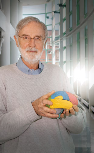
“I think, for better or worse, imaging has become the face of neuroscience.”

Since graduating from medical school in 1964, Raichle has been ...

Interested in reading more?

Become a Member of

The Scientist Logo
Receive full access to digital editions of The Scientist, as well as TS Digest, feature stories, more than 35 years of archives, and much more!
Already a member? Login Here

Related Topics

Meet the Author

  • head shot of blond woman wearing glasses

    Anna Azvolinsky received a PhD in molecular biology in November 2008 from Princeton University. Her graduate research focused on a genome-wide analyses of genomic integrity and DNA replication. She did a one-year post-doctoral fellowship at Memorial Sloan Kettering Cancer Center in New York City and then left academia to pursue science writing. She has been a freelance science writer since 2012, based in New York City.

    View Full Profile

Published In

Share
Illustration of a developing fetus surrounded by a clear fluid with a subtle yellow tinge, representing amniotic fluid.
January 2026, Issue 1

What Is the Amniotic Fluid Composed of?

The liquid world of fetal development provides a rich source of nutrition and protection tailored to meet the needs of the growing fetus.

View this Issue
Skip the Wait for Protein Stability Data with Aunty

Skip the Wait for Protein Stability Data with Aunty

Unchained Labs
Graphic of three DNA helices in various colors

An Automated DNA-to-Data Framework for Production-Scale Sequencing

illumina
Exploring Cellular Organization with Spatial Proteomics

Exploring Cellular Organization with Spatial Proteomics

Abstract illustration of spheres with multiple layers, representing endoderm, ectoderm, and mesoderm derived organoids

Organoid Origins and How to Grow Them

Thermo Fisher Logo

Products

Brandtech Logo

BRANDTECH Scientific Introduces the Transferpette® pro Micropipette: A New Twist on Comfort and Control

Biotium Logo

Biotium Launches GlycoLiner™ Cell Surface Glycoprotein Labeling Kits for Rapid and Selective Cell Surface Imaging

Colorful abstract spiral dot pattern on a black background

Thermo Scientific X and S Series General Purpose Centrifuges

Thermo Fisher Logo
Abstract background with red and blue laser lights

VANTAstar Flexible microplate reader with simplified workflows

BMG LABTECH