Neuroscience

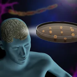
A DNA strand, representing gene therapy, is shown behind a brain.

Gene Therapy Reverses Fragile X Deficits in Mouse Study

Keith Thomas wears a brain-computer interface with black wires and a medical device on his wrist. He is holding hands with researcher Chad Bouton in a research study room.

Paralyzed Man Feels Sensation Again With Brain Stimulation Device

Repairing the Injured Nervous System With Stem Cells

Repairing the Injured Nervous System With Stem Cells

Biotium
Neuron cell anatomy representative of cells that undergo chronoferroptosis.

Long-Term Iron Buildup Weakens Brain Cell Defenses Against Stress

Reconstructed tomography and ultrasound image of the brain’s vasculature and glymphatic system. The blood vessels are visible as weaving strands outlining the shape of a brain, ranging from blue to green to yellow to indicate depth. The cerebrospinal fluid of the glymphatic system appears as bright orange, yellow, and red spots across the top of the brain and, with less intensity, in the middle and at the bottom of the image.

Ultrasound Reveals the Brain’s Waste Removal System

In a black and white photo on the left, Annina Schmid smiles with her arms crossed. In the color-photo on the right she wears a white lab coat and smiles in a research laboratory.

Then and Now: Annina Schmid Uncovers Neuropathic Pain Signatures

Image of clonal raider ants.

Ancient Neuropeptides Shape Modern Ant Parental Care

Two halves of a medical model of a human brain, with the brainstem at the bottom, signifying the structure mapped by ANCHOR brainstem atlas.

A New Brainstem Atlas Reveals Hidden Anatomy of the Human Brain

A drawing shows two scientists colored pink analyzing a brain colored blue.

EBRAINS Crowdsources the Future of European Neuroscience

Photograph of Humsa Venkatesh against a brick wall.

Then and Now: Humsa Venkatesh Decodes How Tumors Hijack the Nervous System

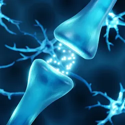
Two neurons communicating with electrical signals

High-Throughput Electrophysiology Workflows for In Vitro Models

Integra Logo

Trending

A team of cartoon scientists studying a giant pancreas.

Prolonged GLP-1 Exposure Remodels Gene Expression in the Pancreas by Flipping a Molecular Switch

A magnifying glass, representing increased scrutiny into this student’s publication record, leans against a stack of papers.

Student Admits She Did Not Conduct Experiments in Now-Retracted Paper

Image of a Diploptera punctata cockroach.

Cockroach Milk Proteins Churn Out an Ig Nobel Win

Illustration of a white piece of 3D DNA against a white background. Cartoon people stand on top of the bases on the bottom strands, representing population-wide genomic studies.

A Meta-Analysis Uncovers Genetic Risk Factors for Fibromyalgia

Multimedia

Rethinking ALS Biomarkers: From Discovery to Clinical Impact

Rethinking ALS Biomarkers: From Discovery to Clinical Impact

Alamar Biosciences logo
Best Practices for qPCR Assay Design and Optimization

Best Practices for qPCR Assay Design and Optimization

Bio-Rad
The Scientist Digest cover September 2026
September 2026

Multiplex Microscopy Becomes Easier with Encoded Antibodies

A new system that enables researchers to uniquely tag monoclonal antibodies for use in microscopy could help simplify complex imaging studies.

View this Issue
Rethinking ALS Biomarkers: From Discovery to Clinical Impact

Rethinking ALS Biomarkers: From Discovery to Clinical Impact

Alamar Biosciences logo
Best Practices for qPCR Assay Design and Optimization

Best Practices for qPCR Assay Design and Optimization

Bio-Rad
Beyond the Basics: Strategies for Single-Cell and Spatial Transcriptomics Analysis

Beyond the Basics: Strategies for Single-Cell and Spatial Transcriptomics Analysis

bioxcell
Scientist reviewing cellular and molecular data on a computer in a laboratory.

Building Translation-Ready Biomarkers with Connected Workflows

Danaher Logo

Products

Closeup image of a multi channel pipette dispensing pink liquid into a 96-well plate.

The ASSIST PLUS pipetting robot for affordable workflow automation

Integra Logo
Single cells in suspension

Rapidly isolate primary cells and make uniform single-cell suspensions with Corning® Cell Strainers

Corning logo
Abstract image representing cell membranes linked together.

CellBrite® Steady Membrane Stain: Cell surface staining built for real-time imaging

Biotium
sino biological logo

Monod Bio Licenses AI-designed Protein Technologies to SignalChem Biotech for Custom Discovery Assays