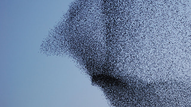
MOTION CONTROL: An immense flock of starlings, known as a murmuration, preparing to roost at dusk© FIONA ELISABETH EXON/GETTY IMAGES

Small silvery schooling fish known as golden shiners are experts at quickly finding shady spots that offer better camouflage from predators. Individual fish flit from one shady spot to another in the ponds and lakes they inhabit, but only appear to sense the changing light when they swim in large schools. When swimming solo, these fish are much less adept at estimating the light levels of their environment. They show little preference for darker areas, suggesting that they have a limited ability, if any at all, to detect the changing brightness of their surroundings.

Such conundrums have always fascinated Iain Couzin, an evolutionary biologist at Princeton University. By observing and tracking the behaviors of golden shiners (Notemigonus crysoleucas) swimming in a pool with varied light levels,...

The higher-order complexity that arises from the compounded actions of many is the cornerstone of collective behavior.

This complex group sensing capability arises from very simple behaviors of individual fish. In large schools, if a few fish out of the group hit upon a darker area, they slow down, which causes them to cluster, much like what happens when a few cars on a busy highway suddenly decelerate. Fish still in the light continue to move quickly, but their social attraction towards their slow neighbors causes the group as a whole to sling into the darker area, slow down, and remain there. Therefore the whole group appears to “sense” and gravitate toward the darkness.

The school acts as a sensor array that becomes more sensitive to light as the number of sensors—or fish—increases. Similar examples of this principle are pervasive in nature, and can be observed in starkly different systems—from huge herds of wildebeest to groups of cells forming tissues, colonies, and biofilms.

A MOLECULAR DANCE: Fluorescently labeled F-actin filaments—each about 5 nm thick and 10 μm long—mounted on myosin motors and provided with a steady supply of ATP, are allowed to walk freely on the surface of a coverslip (shown in schematic, bottom right). At low F-actin concentrations, the filaments move independently from one another, but at concentrations higher than 5 filaments per square micrometer, large-scale patterns such as vortices and waves arise. When the actin crosslinker fascin is added to the assay, dense swirls (large photo) and rotating rings (inset) are observed.COURTESY ANDREAS BAUSCH; PNAS 110:4488-93, 2013

“Individual cells may have a very limited capacity to sense long-range chemical gradients,” Couzin says, but groups of cells, exploiting the same algorithm as golden shiners, may become efficient at sensing their environment over a long range.

This higher-order complexity that arises from the compounded actions of many is the cornerstone of collective behavior. It is a field that has enjoyed a burgeoning interest in the last few decades, as more rigorous experimental and theoretical methods for predicting, tracking, and analyzing the behavior of hundreds, thousands, and even millions of individuals in a group are being developed in disciplines as diverse as mathematics, physics, biology, social sciences, economics, and engineering.

Couzin and other collective behaviorists are attempting to unravel the simple algorithms underlying complex group behaviors to understand not only how ant colonies organize and birds in a flock coordinate their motion, but also more fundamental questions about how these simple rules create novel capabilities in molecular and cellular biological systems.

Small beginnings

While bird flocks, fish schools, and honeybee colonies are among the more iconic examples of collective behavior, the phenomenon is also observed at the molecular level in cellular components.

Actin filaments, which form part of cells’ cytoskeletons and transportation networks, can, under experimental conditions, display collective behaviors reminiscent of living organisms. At high densities, actin filaments “walking” on the surface of a slide (propelled by myosin motors) spontaneously self-arrange into clusters, propagating waves of dense bands and spinning in vortices.

When actin filaments number fewer than five per square micrometer, they parade around randomly, and no large-scale order is observed. But as their concentration increases above that threshold, they begin to form clusters, sometimes up to 500 micrometers in diameter, of similarly aligned filaments. At even higher concentrations—more than 20 filaments per square micrometer—waves of dense bands separated by empty regions appear to propagate throughout the system. Within each band, filaments are tightly packed and aligned in the direction of movement.

These intricate patterns seem to arise from weak and local interactions that are reinforced through repeated collisions. When one filament hits its neighbor, for example, it forces the struck filament to slightly alter its direction to align with the collider. Higher densities increase the frequency of collisions, and through many such collisions, filaments become completely aligned with their neighbors.

The resulting patterns get even more interesting when the actin cross-linking protein, fascin, is added to the mix. In the cell, fascin helps bundle individual strands of actin together to strengthen cytoskeletal structure. When fascin is added in vitro, clusters and bands of pure actin morph into rotating rings and moving streaks of actin-fascin structures.2 (See image above "A Molecular Dance.")

That actin filaments are able to form these complex patterns in vitro in the absence of any genetic or cellular guidance bears testimony to the crucial role that self-organization and collective motion likely play in cellular processes such as cytoskeletal formation and inter- and intracellular transportation networks, says Andreas Bausch, a biophysicist at Technische Universität München who helped develop an actin motility assay for studying collective motion in 2010.3

Taking shape

COLLECTIVE MOVEMENT DURING EMBRYOGENESIS: This image (inset) shows a time capture of the dynamics of endothelial cell movement in developing avian aortas—one of the first large vessels to form in amniote embryos. The cells are moving upstream toward the developing heart. Earlier cell positions are dimmer, actual positions bright. The arrows indicate that cells on the ventral cylinder surface (magenta) move more medially than cells on the dorsal surface (blue)

A HEART IN THE MAKING (inset): Endothelial cells move in a spiral-like pattern during formation of the embryonic heart (large red-green-blue vessels forming the arms of an upside-down Y across the image). The aortas are also visible as blue segments behind the heart and under the inflow.
COURTESY OF ANDRAS CZIROK AND THE KUMC COMPUTATIONAL IMAGING GROUP
Embryogenesis is perhaps the most spectacular example of collective motion at the cellular level. Although the arrangement of tissues and organs that occurs during embryogenesis is in large part orchestrated by well-timed changes in gene expression and downstream changes in biomechanical properties, more spontaneous and self-organized processes characteristic of collective motion also play an important role. (See “Fellow Travelers,” The Scientist, February 2013.)

When the first blood vessels begin to assemble in the embryo of a warm-blooded animal, for example, a few endothelial cells become elongated, file out of the original embryonic cell cluster, and migrate hundreds of micrometers to form cord-like structures known as vascular sprouts that eventually extend and connect with others to form vessels. In the process, these pioneer cells recruit other endothelial cells to do the same.

Just what determines which cells initiate migration is not fully understood, but some researchers think that an extracellular signaling gradient selects the most motile cells in the bunch to act as pioneer cells. Once selected, these cells, in turn, actively suppress the leader-cell properties of their neighbors, explains András Czirók, a cell biologist at the University of Kansas Medical Center. Although the molecules that initiate suppression by pioneer cells haven’t been fully elucidated, a similar inhibition occurs in the developing brain and is responsible for maintaining a subpopulation of neural progenitor cells.4 (See sidebar on left.)

As the pioneer endothelial cells begin to move away from the original cluster of embryonic cells, their newly elongated shape, which extends parallel to the direction of migration, acts as a guidance cue for adjacent cells. During vascular-sprout formation, the ability of leader cells to inhibit neighboring cells is transient, however, and cells that were originally lagging behind become able to take the lead. This process of transient leadership appears throughout the course of collective motion.

Even after large blood vessels, such as the aorta, have formed, the endothelial cells remain highly motile. “We thought that once the blood vessels form, the cells are kind of static, like the bricks of a house,” Czirók says. Instead, the walls of the newly formed aortas of bird embryos revealed a turbulent mosaic of cells, with visible streams and vortices.

Although at first this movement is random, once blood starts pumping through the vessels, the streams of moving cells making up the vessel walls eventually become directed toward completing the still-forming heart. Only a small percentage of the cells—around 5 to 10 percent—are needed to initiate this mass migration of cells into the nascent organ. Rather than heart formation relying on cell division alone, the streaming vascular endothelial cells provide “a fast and easy way to transport new cells into the growing heart,” Czirók says. (See images adjacent.)

Thriving together

IN UNITY, VICTORY: Depending on growth conditions, Paenibacillus dendritiformis bacteria can switch between two distinct morphotypes. When grown on soft surfaces, which allow for greater motion, P. dendritiformis cells become elongated and move in long, slightly curved thin lines. Known as the chiral morphotype, the cells produce colonies with curvy branches that curl in one direction (main image). However, when grown on hard surfaces with limited nutrients, P. dendritiformis switch into the branching, or tip splitting, morphotype (inset), characterized by rounder cells that move in whirls and jets. The branches of the colony formed by this morphotype allow for greater bacterial density, which in turn may result in increased amounts of the cell-secreted lubricant necessary for colony expansion on the hard surface.COURTESY OF ESHEL BEN-JACOBAlthough researchers may have first been inspired by the collective behaviors of macroscopic multicellular organisms such as wildebeests, ants, and schooling fish, some are turning to bacteria, which are easier to track in three-dimensional space over time, and which display behavioral dynamics similar to those of larger animals. “Despite all the beautiful things people observe in birds and locusts and higher animals, if you look at the migration of bacteria, it’s still more sophisticated than any of this,” says Eshel Ben-Jacob, a biological physicist at Tel Aviv University.

In the mid-1990s, Ben-Jacob discovered two strains of bacteria that form some of the most spectacular patterns of collective behavior known. When grown on thin, hard surfaces with limited nutrients, a colony of the soil-dwelling facultative anaerobe Paenibacillus vortex secretes a lubricating fluid that allows the cells to swim and form large colonies with intricate fractal-like branching patterns. At the end of each branch, a swirling vortex of bacteria expands its edges.

Within the swarm, which typically contains a greater number of cells than the number of humans on Earth, lanes and highways much more complex than those formed by shiners or wildebeests transfer information and even cargo throughout the colony.

Ben-Jacob has found that environmental conditions fine-tune epigenetic programming in P. vortex cells. By regulating simple factors, such as the amount and viscosity of fluid they secrete, cells in the collective are able to form large-scale patterns that are advantageous to their circumstances. For example, regulating fluid viscosity—a response that’s dependent on cell density and the availability of nutrients—limits the width of the colony’s branches so that the average density at any given point of the swarm matches the amount of food available in the area. Thus, via individual cell responses to the conditions of the immediate environment, communicated to other cells, the colony as a whole forms remarkably intricate patterns that change along with changing conditions.5 (See images above.)

FACING HUNGER TOGETHER: A starving colony of Myxococcus xanthus forms fruiting bodies. Inset: an image of the edge of an Myxococcus xanthus swarm, where cells are densely packed, but individual cells (about 1 micron wide and 10 microns long) are visible at the outmost edge. TRANCE GEMINI, WIKIMEDIA COMMONS; COURTESY OF YILIN WUSimilarly, the gram-negative species Myxococcus xanthus hunts for food in topsoil as a predatory swarm, but when conditions are unfavorable, some members of the swarm undergo cell differentiation and aggregate into spore-forming fruiting bodies. The complex, ordered states of a Myxococcus swarm are maintained in the absence of any long-range signaling among cells, explains Yilin Wu, a biophysicist now at the Chinese University of Hong Kong.

Instead, by reversing directions every 8 minutes (think of a car reversing and advancing in order to parallel park), cells resolve jams and develop an orientation and alignment both among their immediate neighbors and in relation to cells at a distance. Such orderly alignments within the swarm are crucial for its expansion, and ultimately for the formation of the fruiting body that permits the survival of the colony during tough conditions.6

Because they’re easy to handle experimentally, researchers studying collective behavior are using a number of bacterial species as model organisms, not only to understand basic motility patterns, but ultimately to gain insight into the role of collective movements in biofilm formation during infection and in the transfer of antibiotic resistance among cells. For example, Ben-Jacob has found that the shapes of some bacterial colonies are altered under exposure to antibiotics.7 Although the mechanisms that lead to this alteration (which are presumably epigenetic) remain obscure, it is possible that the new colony structure itself may increase the cells’ ability to survive long enough for them to develop resistance to the antibiotic.

From simple to complex

COLLECTIVE BEHAVIOR AT THE BASAL LEVEL: A representative image for the general amoeboid body plan of the placozoan Trichoplax adhaerens, one of the structurally simplest multicellular animals. The animal moves either by crawling along the surface on a bed of its own cilia, or by extending pseudopod-like projections (1). The placozoan in this image extends a number of these projections. The question of how this animal, which lacks an anterior-posterior axis, decides its direction of motion and establishes a “leading edge” is under active investigation (2). Larger specimens frequently form vertical folds as emergent structures that may be used in asexual reproduction by fission or in environmental sensing (3). In some cases, folds appear highly aligned and form larger patterns across the animal’s entire body (4).COURTESY OF MIRCEA DAVIDESCUColonies of spore-forming bacteria, such as M. xanthus and P. vortex, can reach levels of complexity reminiscent of multicellular systems, containing clusters of differentiated cells that act as reproductive organs, among other things.

“Collective behavior may have been really vital for the evolution of multicellular life on Earth,” says Couzin, who very recently added placozoans—thought to be one of the structurally simplest of all multicellular animals on Earth—to his diverse roster of model organisms.

This basal metazoan can be thought of as a flat collection of around 1,000 to 3,000 cells that scours the oceans for an algal or bacterial meal. The translucent, thin-bodied organisms, about 1 mm in diameter, have proto-neurons and muscle-like cells and share many of the gene pathways found in more advanced animals. Because of the placozoan’s simplicity, “it appears to really need to make collective decisions about many aspects of its life,” says Couzin. “By acting together as a collective, perhaps [the cells] can achieve a degree of intelligence that wouldn’t be possible for individual cells,” Couzin says. Eventually, understanding collective behavior and collective decision making in a primitive multicellular creature such as the placozoan may offer clues about how more-complex multicellular systems first evolved.

A former staff member of  The Scientist, Cristina Luiggi is currently a science writer based in Paris, France.
 

Banding Together

From bacteria to animals—a sampler of organisms that display collective behaviors.


E.COLI
COURTESY OF YILIN WUWhen grown on agar plates under humid and nutrient-rich conditions, Escherichia coli cells elongate and sprout many helical hair-like structures, known as flagella. The bacterial cells also create a thin layer of fluid, a few microns thick, in which they swim by rotating the flagella counterclockwise for propulsion. At high enough densities, E. coli cells align and band together into rafts, forming a swarm. Yilin Wu of the Chinese University of Hong Kong has found that as tens of thousands of cells line the edge of the swarm, the collective action of millions of flagella rotating in a counterclockwise direction generates a “river,” about ten microns wide, that circles the colony. As the colony expands, cells carried out by the current form branches that curve to the right.


MAMMALIAN NEURONS
© AFP/GETTY IMAGESWhen the mammalian brain’s visual cortex is tracking the trajectory of a moving object, neurons will fire with different bits of information gathered from the visual system. Different cell groups compete and inhibit each other until a quorum is reached and the brain makes a collective decision about the direction of movement.


HONEYBEES
CATHEDRAL TERMITE MOUND/WIKIMEDIA COMMONSEvery spring, honeybee queens leave their hives together with several thousand workers to search for new homes. After scoping out potential hive locations, several hundred honeybee scouts return to the swarm and perform intricate waggle dances that convey their choice of location. Encoded in these dances is information about the distance and direction of the selected location, which other bees use to check out and evaluate the quality of the site. In 2011, researchers led by Thomas Seeley of Cornell University found that scout bees performing waggle dances ram their heads against rival scout bees in order to stop them from advertising an alternative location. If a scout bee’s dance is interrupted enough times, it will stop dancing altogether. In the end, the colony chooses the most danced-for location. This inhibition mechanism likely prevents delays in reaching consensus brought on by indecision within the colony.
 

MOUND-BUILDING TERMITES
CATHEDRAL TERMITE MOUND/WIKIMEDIA COMMONSSome termite species, such as Macrotermes subhyalinus, build intricate dirt mounds that can tower more than 30 feet high. These structures serve as homes, nurseries, and farms, and shelter the colony from the fluctuating temperature and humidity of the outside environment. A complex system of tunnels and galleries helps direct air currents throughout the mound, operating both as heating and cooling vents while at the same time helping to minimize carbon dioxide concentrations. Yet, as sophisticated as these termite buildings seem to be, there is no centralized command guiding the building of the nests. Instead, it is self-organized by individual termites responding to simple environmental cues such as temperature, humidity, carbon dioxide levels, air flow, and pheromone trails laid down by nest mates.
 

HOPPERS
© WALTER GALLOWAY/ISTOCKPHOTO.COMUnlike ants or bees, which gather together out of the need for cooperation, certain swarms of insects, such as the desert locust (Schistocerca gregaria, pictured above) and the Mormon cricket (Anabrus simplex), counterintuitively form large bands of marching individuals in order to avoid being cannibalized by their own. As critical nutrients such as protein and salt run low, and the density of wingless juveniles increases, individuals begin to turn on each other. Insects grouped together will try to eat those in front, while at the same time avoiding being cannibalized from behind, until eventually they arrange themselves into orderly files. “The outcome is a vast swarm that is moving collectively across the environment,” says Princeton’s Iain Couzin, who discovered the cannibalistic origins of locust swarms. “It’s a forced march.”
 

STARLINGS
COURTESY OF SILVIO DUARTE QUEIROSAlthough flocking starlings (Sturnus vulgaris) have fascinated birdwatchers for millennia with their complex formations and fluid movements, recent research into the 3-dimensional distribution of starling flocks has revealed that the shape of the flock is surprisingly flat. Despite the variety of shapes they form, the birds are not as distributed vertically as they are sideways. The coherent movements of the flock as a whole are maintained because the individual starlings continually align themselves to their neighbors, referencing only the six or seven birds closest to them. However, over time the birds will drift to gain new neighbors. As a result they can coordinate their motion even more efficiently. In fact, information, such as changes in speed and direction, travels throughout a flock of starlings almost instantly—with birds at one edge of the flock responding to the behavior of birds at the other edge almost simultaneously. These scale-free behavioral correlations are maintained regardless of flock size.

References

  1. A. Berdahl et al., “Emergent Sensing of Complex Environments by Mobile Animal Groups,”
    Science, 339:574-56, 2013.
  2. V. Schaller et al., “Frozen steady states in active systems,” PNAS, 108:19183-88, 2011.
  3. V. Schaller et al., “Polar patterns of driven filaments,” Nature, 467:73-77, 2010.
  4. E.D. Perryn et al., “Vascular sprout formation entails tissue deformations and VE-cadherin-dependent cell-autonomous motility,” Dev Biol, 313:545-55, 2008.
  5. A. Sirota-Madi et al., “Genome sequence of the pattern forming Paenibacillus vortex bacterium reveals potential for thriving in complex environments,” BMC Genomics,
    11:710, doi:10.1186/1471-2164-11-710, 2010.
  6. Y. Wu et al., “Periodic reversal of direction allows Myxobacteria to swarm,” PNAS, 106:1222-27, 2009.
  7. E. Ben-Jacob et al., “Bacterial cooperative organization under antibiotic stress,” Physica A: Statistical Mechanics and its Applications, 282:247-282, 2000.

Interested in reading more?

Magaizne Cover

Become a Member of

Receive full access to digital editions of The Scientist, as well as TS Digest, feature stories, more than 35 years of archives, and much more!