Eyes grown from stem cells

Cultured mouse embryonic stem cells self-organize into a complex retinal structure

| 3 min read

Register for free to listen to this article
Listen with Speechify
0:00
3:00
Share
With the proper culture conditions, mouse embryonic stem (ES) cells can spontaneously form the rudiments of a retina -- the neural tissue and most complex component of the eye. The results, published today (April 6) in Nature, could help researchers answer some outstanding questions about eye development and dysfunction, and hold promise for the development of retinal tissues for transplantation.
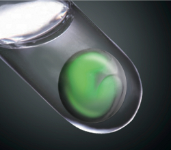
A conceptual image of an ES cell-derived optic cup in a test tube.
Image: M. Eiraku and Y.Sasai at RIKEN Center for Developmental Biology
"I haven't been so excited about a study in a very long time," said retinal geneticist linkurl:Robin Ali;http://www.ucl.ac.uk/ioo/pdf/PI/Professor%20Robin%20Ali.pdf of University College London, who was not involved in the research. "Until now, it's not been possible to develop organs or complex tissues from stem cells.""That's incredible to me that you can do this in vitro," agreed developmental biologist linkurl:Panagiotis Tsonis;http://udbiology.com/content.php?id=1131 of the Center for Tissue Regeneration and Engineering at the University of Dayton, who did not participate in the study. "I think this is one of the best papers I've seen in the stem cell field."For the last decade, developmental biologist linkurl:Yoshiki Sasai;http://www.cdb.riken.jp/en/02_research/0201_core05.html at the RIKEN Center for Developmental Biology in Japan and his colleagues have worked to differentiate ES cells into various cells of the nervous system, including cerebral cortex neurons and retinal cells. But Sasai was interested in more than just generating different nerve cells. He wanted to learn how those cells come together to form entire tissues and organs in developing embryos.Starting with the culture conditions they had established for retinal differentiation, the researchers added matrix proteins that they hoped would encourage the formation of the more rigid retinal epithelial structures. They then seeded the culture with mouse ES cells. Within a week, the cells began to form small vesicles and differentiate into two different tissue types: Cells on one side of the vesicles formed the mechanically rigid pigment epithelium, while cells on the other side differentiated into a more flexible tissue that folded inward in the shape of an embryonic optic cup -- the retina's precursor.
Dynamic formation of an optic cup in 3D culture of an ES cell aggregate.
Green represents retinal precursor tissue.
Video from M. Eiraku and Y. Sasai at RIKEN Center for
Developmental Biology
"The biggest surprise was that we observed the formation of the very real optic cup structure that mimicked both the shape and tissue composition and popped out from the [ES cell] aggregate," Sasai told The Scientist in an email. "It is an emergence of complex pattern from no pattern."The generation of retinal tissue from ES cells is an exciting advance that may lead to regenerative medicine applications. While doctors are not about to start transplanting these synthetic retinas, Ali said, ES cells cultured under the proper conditions could yield certain cells that may prove therapeutically valuable. "What this would do is provide a source of cells that we could use [for transplantation]," said Ali, who coauthored an accompanying perspective article in Nature. "I think it's a big landmark for work in developing retinal cell therapies."
Two ES cell-derived optic cup formed by self-organization in 3D culture. Green color is fluorescence of GFP protein that was engineered to mark retinal tissue.
Image: M. Eiraku and Y.Sasai at RIKEN Center for Developmental Biology
The results may also be a boon to basic biology, as researchers can now image and watch the development of a retina in real time. For example, the system might help answer how ES cells self-organize into the complex retinal tissues. "Certain mechanical forces were involved to shape the cup," Tsonis said, but the details of that process are still unclear. While the retinal structures cultured in this study only developed into neonatal mouse retinas, which still lack photoreceptor cells, Ali said, it will likely just take a few tweaks to the culture conditions to coax those structures into mature retinas, allowing researchers to examine the entire process. Furthermore, if researchers can replicate the results using human induced pluripotent stem (iPS) cells, it could shed light on retinal dysfunction, Ali added. "By creating iPS cells from patients with [visual disorders] and then making synthetic retinas from such iPS cells, we could potentially study the disease process caused by particular genetic defects."However the system is applied, there is no doubt about the study's significance. "It truly is stunning," Ali said. "I never thought that I'd ever see a retina grown in a dish."M. Eiraku et al., "Self-organizing optic-cup morphogenesis in three-dimensional culture," Nature, 472: 51-6, 2011.
**__Related stories:__***linkurl:Eye evolution questioned;http://www.the-scientist.com/news/display/58032/
[1st March 2011]*linkurl:New master switch in brain?;http://www.the-scientist.com/blog/display/57542/
[1st July 2010]*linkurl:Let's grow organs;http://www.the-scientist.com/blog/display/54171/
[15th January 2008]
Interested in reading more?

Become a Member of

The Scientist Logo
Receive full access to more than 35 years of archives, as well as TS Digest, digital editions of The Scientist, feature stories, and much more!
Already a member? Login Here

Keywords

Meet the Author

  • Jef Akst

    Jef Akst was managing editor of The Scientist, where she started as an intern in 2009 after receiving a master’s degree from Indiana University in April 2009 studying the mating behavior of seahorses.
Share
May digest 2025 cover
May 2025, Issue 1

Study Confirms Safety of Genetically Modified T Cells

A long-term study of nearly 800 patients demonstrated a strong safety profile for T cells engineered with viral vectors.

View this Issue
Detecting Residual Cell Line-Derived DNA with Droplet Digital PCR

Detecting Residual Cell Line-Derived DNA with Droplet Digital PCR

Bio-Rad
How technology makes PCR instruments easier to use.

Making Real-Time PCR More Straightforward

Thermo Fisher Logo
Characterizing Immune Memory to COVID-19 Vaccination

Characterizing Immune Memory to COVID-19 Vaccination

10X Genomics
Optimize PCR assays with true linear temperature gradients

Applied Biosystems™ VeriFlex™ System: True Temperature Control for PCR Protocols

Thermo Fisher Logo

Products

The Scientist Placeholder Image

Biotium Launches New Phalloidin Conjugates with Extended F-actin Staining Stability for Greater Imaging Flexibility

Leica Microsystems Logo

Latest AI software simplifies image analysis and speeds up insights for scientists

BioSkryb Genomics Logo

BioSkryb Genomics and Tecan introduce a single-cell multiomics workflow for sequencing-ready libraries in under ten hours

iStock

Agilent BioTek Cytation C10 Confocal Imaging Reader

agilent technologies logo