
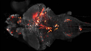
 Brain of a zebrafish larva showing near-simultaneous activation of a large population of neurons (in red). MISHA B. ARENS AND PHILIPP J. KELLERResearchers have recorded and visualized the activity of nearly every individual neuron in the larval zebrafish brain in real time, according to a study published this week (March 18) in Nature Methods. The technique should help scientists to better understand how local circuits of neurons form large-scale networks across different brain regions.
“It allows a much better view of the dynamics throughout the brain during different behaviors and during learning paradigms,” Joseph Fetcho, a neurobiologist at Cornell University in New York, told Nature.
Last year, Misha Ahrens and Philipp Keller, neurobiologists at the Janelia Farm Research Campus in Ashburn, Virginia, imaged the brain activity of zebrafish larvae as they navigated through a virtual reality environment. The researchers used light-sheet photography, in which thin sheets of light illuminate the sample and a detector captures images at repeated intervals, to view of the brains of genetically engineered zebrafish in which neurons express a calcium ion-sensing protein that fluoresces when those neurons fire.
For the latest study, the researchers have improved the system to record the activity of a greater ...


