Fruit-Fly Neurons in Action

Researchers visualize the complete nervous system of a Drosophila melanogaster larva at nearly single-neuron resolution.

Written byJef Akst
| 2 min read

Register for free to listen to this article
Listen with Speechify
0:00
2:00
Share

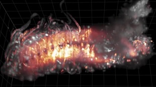
Fruit fly nervous system in actionYOUTUBE, SCINEWSA new video reveals flashes of neural activity as it moves up and down the body of a fruit-fly larva. The feat, published yesterday (August 11) in Nature Communications, adds Drosophila melanogaster to the list of species—including C. elegans and larval zebrafish—whose nervous systems have been imaged at single-cell or nearly single-cell resolution.

“The new thing we can do now is study how the neurons talk to each other across the entire brain,” Misha Ahrens, a neuroscientist at the Howard Hughes Medical Institute’s Janelia Research Campus in Ashburn, Virginia, who worked on the zebrafish study, told The Scientist in February 2014. “That’s a departure from previous studies, where people tended to look at many neurons, but a small fraction of the entire number.”

The new study builds on Ahrens and his colleagues’ method of genetically modifying neurons to fluoresce as they fire, then using a microscopy technique to shoot light at the brain and record its activity. The resulting video shows the activity of the entire Drosophila-larva central nervous system, which was stripped from the body for better ...

Interested in reading more?

Become a Member of

The Scientist Logo
Receive full access to more than 35 years of archives, as well as TS Digest, digital editions of The Scientist, feature stories, and much more!
Already a member? Login Here

Related Topics

Meet the Author

  • Jef (an unusual nickname for Jennifer) got her master’s degree from Indiana University in April 2009 studying the mating behavior of seahorses. After four years of diving off the Gulf Coast of Tampa and performing behavioral experiments at the Tennessee Aquarium in Chattanooga, she left research to pursue a career in science writing. As The Scientist's managing editor, Jef edited features and oversaw the production of the TS Digest and quarterly print magazine. In 2022, her feature on uterus transplantation earned first place in the trade category of the Awards for Excellence in Health Care Journalism. She is a member of the National Association of Science Writers.

    View Full Profile
Share
Illustration of a developing fetus surrounded by a clear fluid with a subtle yellow tinge, representing amniotic fluid.
January 2026

What Is the Amniotic Fluid Composed of?

The liquid world of fetal development provides a rich source of nutrition and protection tailored to meet the needs of the growing fetus.

View this Issue
Redefining Immunology Through Advanced Technologies

Redefining Immunology Through Advanced Technologies

Ensuring Regulatory Compliance in AAV Manufacturing with Analytical Ultracentrifugation

Ensuring Regulatory Compliance in AAV Manufacturing with Analytical Ultracentrifugation

Beckman Coulter Logo
Skip the Wait for Protein Stability Data with Aunty

Skip the Wait for Protein Stability Data with Aunty

Unchained Labs
Graphic of three DNA helices in various colors

An Automated DNA-to-Data Framework for Production-Scale Sequencing

illumina

Products

nuclera logo

Nuclera eProtein Discovery System installed at leading Universities in Taiwan

Brandtech Logo

BRANDTECH Scientific Introduces the Transferpette® pro Micropipette: A New Twist on Comfort and Control

Biotium Logo

Biotium Launches GlycoLiner™ Cell Surface Glycoprotein Labeling Kits for Rapid and Selective Cell Surface Imaging

Colorful abstract spiral dot pattern on a black background

Thermo Scientific X and S Series General Purpose Centrifuges

Thermo Fisher Logo