
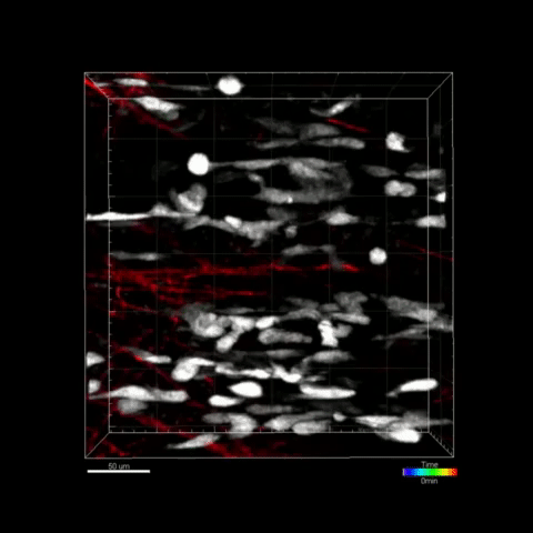
 Activated muscle stem/progenitor cells (white) moving within basal lamina remnants (ghost fibers in red)CARNEGIE INSTITUTE FOR SCIENCE: MICAH WEBSTER, CHEN-MING FAN; NICHD: JENNIFER LIPPINCOTT-SCHWARTZ, URI MANORAt the site of a muscle injury, the dying muscle fibers leave behind “ghost fibers,” remnants of the extracellular matrix. According to a study published by researchers from the Carnegie Institution for Science and the National Institute of Child Health and Human Development last week (December 10) in Cell Stem Cell, these fibers help guide new muscle cells to grow in place to heal the injury in mice.
The research team anesthetized a set of mice and then either injected the animals’ hind limb muscles with a cardiotoxin or made manual incisions into the muscle. The striated muscle tissue began disappearing from wound sites one day after injury, leaving behind a series of collagen ghost fibers.
Using two-photon imaging and second-harmonic generation microscopy, the team was able to observe the stem cells and muscle precursor cells in each mouse orienting themselves along the ghost fibers as they prepared to grow new muscle tissue. When the researchers perturbed the system and changed the orientation of the ghost fibers, the resulting muscle tissue grew back improperly.
In their paper, the authors proposed that “the ghost fiber (1) is a key determinant for patterning muscle stem ...


