 Infographic: Growth Plate Dynamics
View full size JPG | PDF LUCY READING - IKKANDA
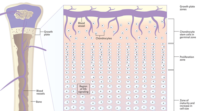
The growth plate is divided into several distinct regions, each populated with cartilage cells (chondrocytes) displaying characteristic behaviors. Nearest the heads of the bone is a germinal zone containing stem cells, below which is a zone of proliferating chondrocytes, followed by one in which the chondrocytes develop into orderly columns of cartilage and the cells increase in size from 4- to 10-fold. At the bottom end of the growth plate, the chondrocytes undergo programmed cell death and are replaced by bone. What determines the size of these regions is not known. The number of cells in a column can be as high as 40, and cells can be produced at rates of over 10,000 a day. This huge number of cells must be roughly the ...


