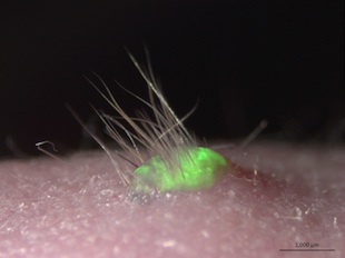
 Transplantation of the bioengineered, 3D integumentary organ system using mouse iPS cells labeled with GFP© RIKEN, TAKASHI TSUJIScientists just got one step closer to growing functional skin in vitro. A team at Japan’s Riken Centre for Developmental Biology has grown skin from mouse induced pluripotent stem cells (iPSCs) that sprouted hair and developed sweat glands when implanted into living animals, according to a paper published in Science Advances last week (April 1).
With this new technique, we have successfully grown skin that replicates the function of normal tissue,” study coauthor Takashi Tsuji of Riken told BBC News. “We are coming ever closer to the dream of being able to recreate actual organs in the lab for transplantation.”
It’s not the first attempt to grow skin in the lab. In 2014, researchers in the UK grew human skin epidermis from stem cells. But the skin produced by these efforts lacked essential features like hair follicles and sweat glands, according to BBC News.
For the present study, Tsuji and his colleagues took cells from the gums of mice and converted them into iPSCs. They fluorescently labeled the cells and grew them in vitro to form tissue with three skin ...