Hearing Channel Components Mapped

Localization of two proteins important for inner ear hair cell function suggests they are part of the elusive mechanotransduction channel.

Written byKerry Grens
| 2 min read

Register for free to listen to this article
Listen with Speechify
0:00
2:00
Share

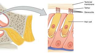
WIKIMEDIA, OPENSTAX COLLEGEHair cells of the inner ear turn sound into electrical nerve signals through a so-called mechanotransduction channel, whose identity remains under wraps. A number of proteins have been implicated as part of this mysterious complex. Researchers have now reported that two of the suspects, TMC1 and TMC2, localize to just the right spots on hair cell tips.

The team generated mice that expressed fluorescently tagged versions of the Tmc genes. In the cochlea, hair cells have finger-like shoots arranged in stadium-seating rows. During the animals’ early development, the fluorescence showed up along the length of these shoots, called stereocilia, and at the tips. Later in life, the proteins were most abundant at the tips of the lower rows of stereocilia. TMC1 remains at the tips through adulthood, whereas TMC2 disappears.

All of this falls in line with the idea that TMC is part of the mechanotransduction channel. The finding that TMC1 and TMC2 are not present at the top of the tallest row of stereocilia, for instance, is consistent with findings that mechanotransduction doesn’t occur there.

“In our current study localizing TMC1 ...

Interested in reading more?

Become a Member of

The Scientist Logo
Receive full access to more than 35 years of archives, as well as TS Digest, digital editions of The Scientist, feature stories, and much more!
Already a member? Login Here

Related Topics

Meet the Author

  • kerry grens

    Kerry served as The Scientist’s news director until 2021. Before joining The Scientist in 2013, she was a stringer for Reuters Health, the senior health and science reporter at WHYY in Philadelphia, and the health and science reporter at New Hampshire Public Radio. Kerry got her start in journalism as a AAAS Mass Media fellow at KUNC in Colorado. She has a master’s in biological sciences from Stanford University and a biology degree from Loyola University Chicago.

    View Full Profile
Share
December digest cover image of a wooden sculpture comprised of multiple wooden neurons that form a seahorse.
December 2025, Issue 1

Wooden Neurons: An Artistic Vision of the Brain

A neurobiologist, who loves the morphology of cells, turns these shapes into works of art made from wood.

View this Issue
Stacks of cell culture dishes, plates, and flasks with pink cell culture medium on a white background.

Driving Innovation with Cell Culture Essentials

Merck
Stacks of cell culture dishes, plates, and flasks with pink cell culture medium on a white background.

Driving Innovation with Cell Culture Essentials

MilliporeSigma purple logo
Human iPSC-derived Models for Brain Disease Research

Human iPSC-derived Models for Neurodegenerative Disease Research

Fujifilm
Abstract wireframe sphere with colorful dots and connecting lines representing the complex cellular and molecular interactions within the tumor microenvironment.

Exploring the Inflammatory Tumor Microenvironment 

Cellecta logo

Products

Beckman Logo

Beckman Coulter Life Sciences Introduces the Biomek i3 Benchtop Liquid Handler, a Small but Mighty Addition to its Portfolio of Automated Workstations

brandtech logo

BRANDTECH® Scientific Announces Strategic Partnership with Copia Scientific to Strengthen Sales and Service of the BRAND® Liquid Handling Station (LHS) 

Top Innovations 2026 Contest Image

Enter Our 2026 Top Innovations Contest

Biotium Logo

Biotium Expands Tyramide Signal Amplification Portfolio with Brighter and More Stable Dyes for Enhanced Spatial Imaging