Improving Zika Virus Detection in Infants

Head circumference is not an accurate indicator of viral infection in newborns, researchers report.

Written byCatherine Offord
| 2 min read

Register for free to listen to this article
Listen with Speechify
0:00
2:00
Share

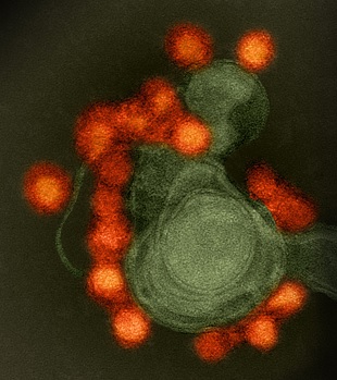
Zika virus (red) isolated from a microcephaly case in BrazilFLICKR, NIAIDZika virus has been linked to microcephaly in the babies of infected mothers, leading some organizations, including the Brazilian ministry of health, to focus on newborns’ head circumferences as an infection indicator. But this approach only identifies a fraction of Zika cases in newborns, researchers reported yesterday (June 29) in The Lancet, suggesting that measurements of head circumference should be combined with other criteria to provide a more accurate diagnosis.

“One in five definite or probable Zika cases had head circumference values in the normal range,” study coauthor Cesar Victora of the Federal University of Pelotas in Brazil said in a statement. “Therefore, the current focus on microcephaly screening alone is too narrow.”

In their review of 1,501 suspected cases of microcephaly investigated in Brazil between November 2015 and February 2016, the researchers also found that rashes—a potential indicator of infection—reported in mothers late in pregnancy were associated with brain abnormalities in newborns even if the babies’ head circumferences were in the normal range. “We should not equate Zika congenital infection with microcephaly,” Victora told CBC News. “We could well have many babies with normal head size who are affected. We will need to think ...

Interested in reading more?

Become a Member of

The Scientist Logo
Receive full access to more than 35 years of archives, as well as TS Digest, digital editions of The Scientist, feature stories, and much more!
Already a member? Login Here

Related Topics

Meet the Author

  • After undergraduate research with spiders at the University of Oxford and graduate research with ants at Princeton University, Catherine left arthropods and academia to become a science journalist. She has worked in various guises at The Scientist since 2016. As Senior Editor, she wrote articles for the online and print publications, and edited the magazine’s Notebook, Careers, and Bio Business sections. She reports on subjects ranging from cellular and molecular biology to research misconduct and science policy. Find more of her work at her website.

    View Full Profile
Share
Illustration of a developing fetus surrounded by a clear fluid with a subtle yellow tinge, representing amniotic fluid.
January 2026, Issue 1

What Is the Amniotic Fluid Composed of?

The liquid world of fetal development provides a rich source of nutrition and protection tailored to meet the needs of the growing fetus.

View this Issue
Skip the Wait for Protein Stability Data with Aunty

Skip the Wait for Protein Stability Data with Aunty

Unchained Labs
Graphic of three DNA helices in various colors

An Automated DNA-to-Data Framework for Production-Scale Sequencing

illumina
Exploring Cellular Organization with Spatial Proteomics

Exploring Cellular Organization with Spatial Proteomics

Abstract illustration of spheres with multiple layers, representing endoderm, ectoderm, and mesoderm derived organoids

Organoid Origins and How to Grow Them

Thermo Fisher Logo

Products

Brandtech Logo

BRANDTECH Scientific Introduces the Transferpette® pro Micropipette: A New Twist on Comfort and Control

Biotium Logo

Biotium Launches GlycoLiner™ Cell Surface Glycoprotein Labeling Kits for Rapid and Selective Cell Surface Imaging

Colorful abstract spiral dot pattern on a black background

Thermo Scientific X and S Series General Purpose Centrifuges

Thermo Fisher Logo
Abstract background with red and blue laser lights

VANTAstar Flexible microplate reader with simplified workflows

BMG LABTECH